Antecedent Surgical Repair Post-Ductal Coarctation with Persistent Mild Stenosis; Subvalvular Aortic Stenosis
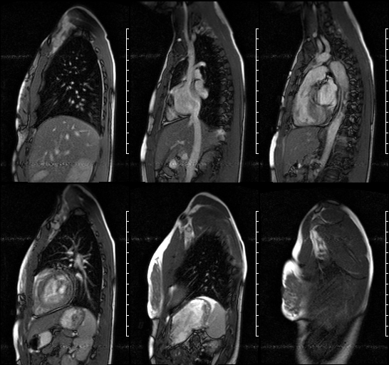
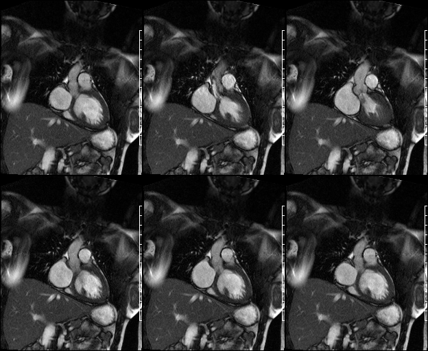
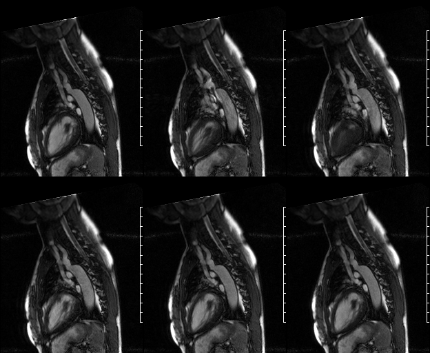
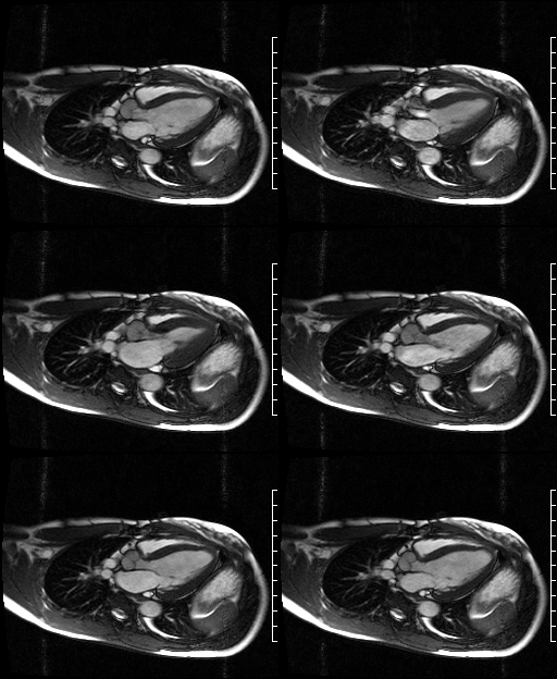
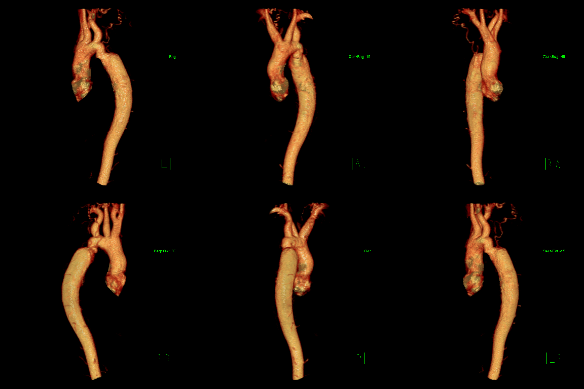
Radiologic Findings Selected cardiac MRI images. Sagittal SSFP (Fig. 1) reveals a focal contour abnormality and region of stenosis involving the proximal descending thoracic aorta just distal to the left subclavian aorta take-off. This is seen to better advantage on the accompanying sagittal oblique SSFP cine targeted to this region of concern (Fig. 3) and on the accompanying 3-D volume rendered MRA of the thoracic aorta (Fig. 5). The latter also reveals recruitment of numerous collateral vessels distal to the point of focal narrowing in the thoracic aorta. Note the turbulent blood flow across the stenotic region in the proximal descending aorta (Fig. 3). Subsequent velocity encoded sequences and quantitative analysis showed a gradient of 30mmHg. Coronal (Fig. 2) and 3-chamber (Fig. 4) SSFP cine images demonstrate mild aortic insufficiency and an area of subvalvular membranous aortic stenosis resulting in a calculated peak gradient of 40mmHg. Note the thin subvalvular membrane anterior to the point of maximum stenosis. Small field-of-view images targeted to the aortic valve (not illustrated) revealed a trileaflet valve with mild failure of central cooptation. Additional imaging findings included a hyperdynamic left ventricle with concentric hypertrophy and a persistent left-sided superior vena cava (Images courtesy of Jennifer Hubert, MD, VCU Medical Center). Diagnosis: Antecedent Surgical Repair Post-Ductal Coarctation with Persistent Mild Stenosis; Subvalvular Aortic Stenosis
Differential Diagnosis
None
Discussion
Coarctation of the thoracic aorta is a congenital anomaly of unknown etiology that results in a focal obstruction at the junction of the distal aortic arch and descending aorta near the ligamentum arteriosum. It accounts for approximately 6% of congenital heart anomalies. Although there is blood flow across the obstruction, it is supplemented by collateral flow that bypasses the obstructing segment via the intercostal arteries, spinal artery, lateral thoracic artery, periscapular arteries, and epigastric arteries.
Pseudocoarctation of the thoracic aorta represents a “kink” in the descending thoracic aorta at the ligamentum arteriosum without obstruction, pressure gradient, or collateral blood flow. It results from elongation of the ascending aorta.
Aortic Stenosis may be acquired (e.g., rheumatic heart disease (usually associated with mitral valve disease); fibrocalcific (senile or degenerative) or congenital (more common). Congenital aortic stenosis may be supravalvular, valvular, or subvalvular. Supravalvular aortic stenosis may be isolated but is also seen in association with Marfan and William syndrome and most often manifests as a localized “hourglass” narrowing just above the aortic sinuses. Valvular aortic stenosis may also occur in isolation but is also associated with coarctation of the aorta. It most often results from fusion of the commissures between the valvular cusps resulting in a bicuspid, unicuspid, or dysplastic valve. Subvalvular aortic stenosis may be either functional (e.g., hypertrophic obstructive cardiomyopathy) or anatomic (i.e., progresses from a thin subvalvular membrane as in this case, to an irregular fibromuscular band, to a thickened left ventricular outflow tract). The anatomic form of subvalvular aortic stenosis is often associated with concomitant congenital heart disease, most commonly ventricular septal defects, patent ductus arteriosus, and aortic coarctation.
Clinical Findings
Isolated aortic coarctation more commonly occurs in men (2.5M: 1F). Coarctation associated with congenital cardiac anomalies affects men and women equally. Associated conditions include Turner syndrome, bicuspid aortic valve, mitral valve prolapse, patent ductus arteriosus, and ventricular septal defects. Patients with isolated coarctation may be entirely asymptomatic. Others become symptomatic latter in life often presenting with headache, claudication fatigue, differential blood flow or pressures between the upper and lower extremities or aortic dissection. Patients with associated cardiac anomalies become symptomatic at birth and often present with left heart failure. Aortic stenosis results in an increased pressure gradient across the aortic valve which eventually causes left ventricular hypertrophy. The coronary vessels do not adequately perfuse the increased left ventricular muscle mass which results in subendocardial ischemia, angina, and left ventricular decompensation with pulmonary venous hypertension. Severe stenosis may be associated with sudden death.
Treatment
Coarctation
- Resection of the coarct segment and graft replacement
- Balloon dilatation of the coarctation for poor surgical candidates
Subvalvular Aortic Stenosis
- Progressive disorder
- Intervention often required at some point in clinical course to relieve left ventricular outflow tract obstruction
- In some patients, progressive aortic regurgitation requires aortic valve repair or replacement at time of surgical intervention
- Percutaneous balloon dilation of discrete subvalvular aortic stenosis can briefly reduce left ventricular outflow pressure gradient
- Current surgical practices are individualized to the specific patient but may entail aortoventriculoplasty in combination with aortic root replacement using a prosthetic aortic valve, an aortic valve allograft, or a pulmonary valve autograft
Prognosis
Coarctation
- Good with early surgical treatment
- Late surgical complications include aneurysm; pseudoaneurysm; infective endocarditis; re-coarctation
Subvalvular Aortic Stenosis
- Progressive disorder
- 2-10% of sudden deaths occur in untreated individuals with severe left ventricular outflow tract obstruction, including subvalvular aortic stenosis, valvular aortic stenosis, and supravalvular aortic stenosis
- Aortic regurgitation eventually develops patients (65%)
- Subvalvular aortic stenosis may recur even after surgical resection
Selected Readings
- Gersony WM. Natural History of Discrete Subvalvular Aortic Stenosis: Management Implications. J Am Coll Cardiol 2001; 38(3):843-845.
- [Guideline] Bonow RO, Carabello BA, Kanu C, et al. ACC/AHA 2006 Guidelines for the Management of Patients with Valvular Heart Disease: A Report of the American College of Cardiology/American Heart Association Task Force on Practice Guidelines (writing committee to revise the 1998 Guidelines for the Management of Patients With Valvular Heart Disease): Developed in Collaboration with the Society of Cardiovascular Anesthesiologists: Endorsed by the Society for Cardiovascular Angiography and Interventions and the Society of Thoracic Surgeons. Circulation 2006; 114(5):e84-231.
- Parker MS, Rosado de-Christenson ML, Abbott GF. Coarctation of the Aorta. In: Teaching Atlas of Chest Imaging 2006; Thieme, New York; 53-57.
- Steiner RM. Reddy GP, Flicker S. Congenital cardiovascular Disease in the Adult Patient: Imaging Update. J Thorac Imaging 2002; 17: 1-17.
Original case written by its authors at Virginia Commonwealth University and published at this address as part of a weekly teaching collection. Reproduced here as an archive.