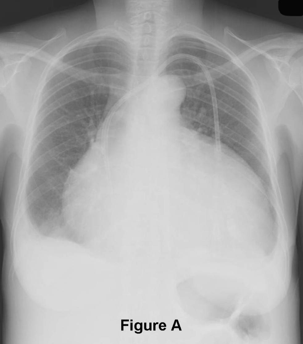
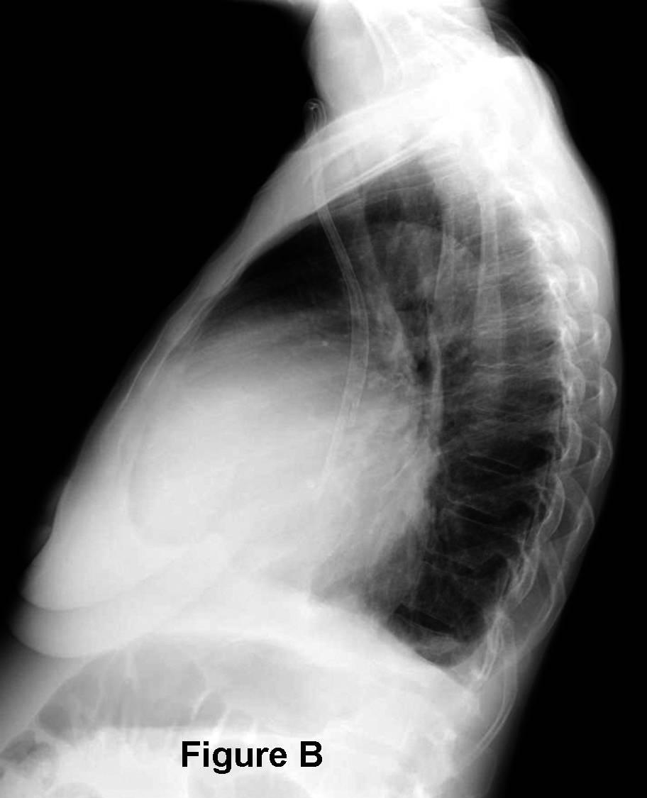
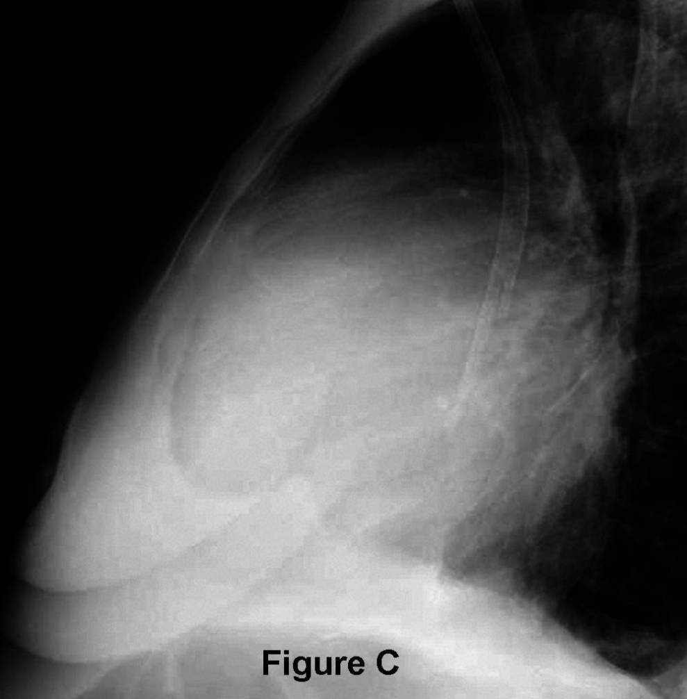
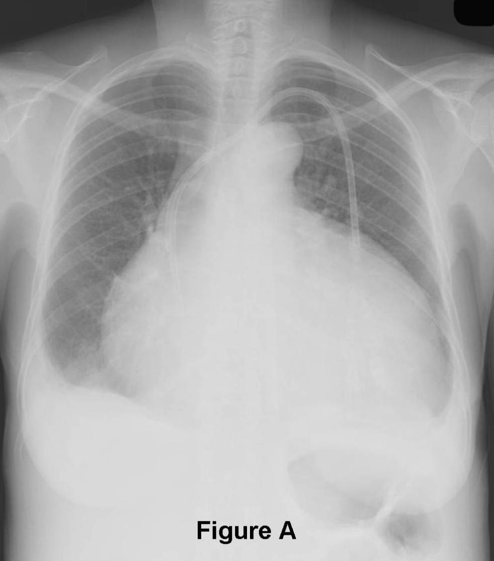
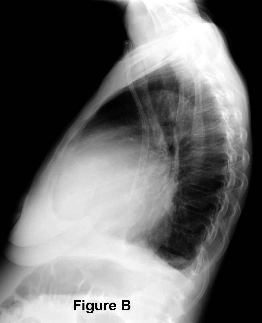
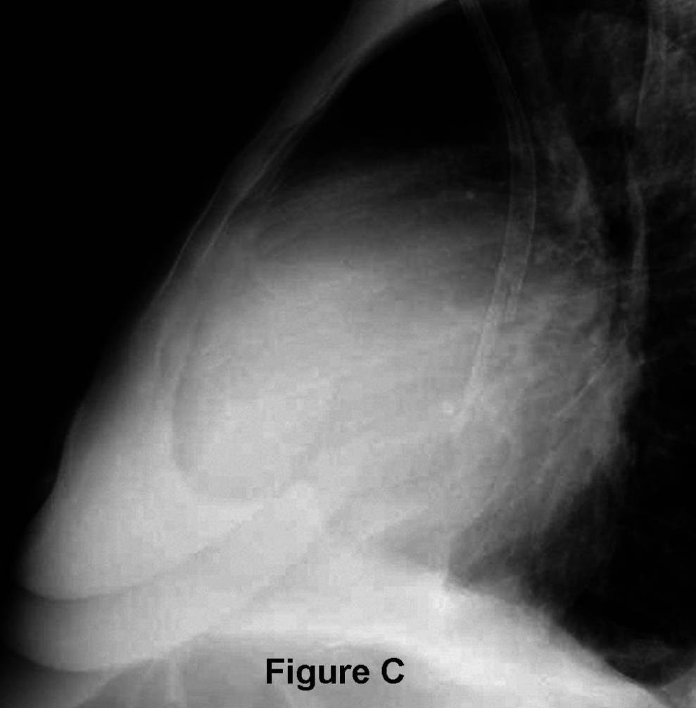
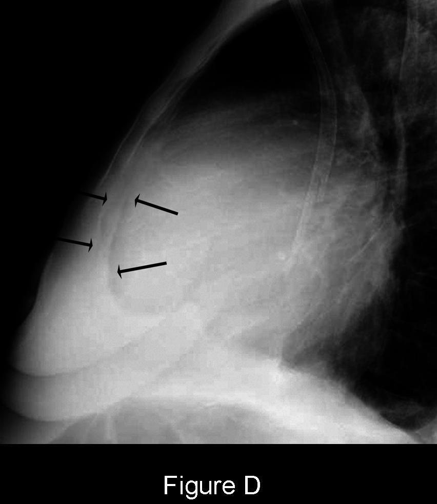
PA (Fig. A) chest x-ray reveals an enlarged cardiomediastinal silhouette with an increase in the transverse diameter but no increase in its height and straightening of the upper mediastinal borders creating a globular or water bottle morphology (“water bottle” sign). The vascular clarity is slightly diminished and bilateral pleural effusions are present, right greater than left. Lateral chest exam (Fig. B) also shows globular enlargement of the cardiomediastinal silhouette and separation of the outer retrosternal and inner epicardial fat lines (“fat pad” or “Oreo? cookie” sign). This later sign is seen to better advantage on the coned-down lateral view (Fig. C and D) (arrows). Diagnosis: Pericardial Effusion
Differential Diagnosis
- Global cardiomegaly-cardiomyopathy
Discussion
The pericardium consists of two layers. Visceral pericardium is attached to the surface of the heart and the proximal great vessels. Parietal pericardium forms the free wall of the pericardial sac. The sac normally contains 20-50 ml of fluid. The most common cause of pericardial effusion is myocardial infarction with left ventricular failure. 50% of patients with chronic renal failure develop uremic pericarditis. Coxsackievirus group B, Staphylococcus, and Hemophilus influenza are common infectious agents associated with pericardial effusion. Today, tuberculous pericarditis is unusual except in the HIV-AIDS population in which pericardial effusion of any etiology is a poor prognostic sign.
Etiology
Increased volumes of pericardial fluid and alterations in the composition of normal pericardial fluid may occur in the setting of numerous diseases (Table -1).
Table-1. Causes of Pericardial Effusion
Serous Hemorrhagic Fibrinous Chylous Heart Failure Acute Infarction Infections Congenital Hypoalbuminemia Cardiac Surgery Uremia SVC Obstruction Irradiation Trauma Rheumatoid Arthritis Cardiothoracic Surgery Myxedema Coagulopathy Systemic Lupus Erythematosus Neoplasia Drug Reactions Neoplasia Hypersensitivity Reaction Thoracic Duct Injuries Aortic Dissection HIV-AIDS
Clinical Findings
Patients may have a relatively large pericardial effusion and experience little or no clinical signs or symptoms, particularly if the fluid has increased slowly over time. This more commonly occur when the pericardial effusion is the result of neoplastic disease or a chronic inflammatory disorder (e.g., rheumatoid arthritis). Alternatively, patients with pericardial effusion may experience dyspnea, orthopnea, both pleuritic and non-pleuritic chest pain, cough, syncope or near syncope, fatigue, tachycardia, and low-grade fever. Tamponade occurs when the volume of fluid in the pericardial sac compromises blood return to the right heart affecting cardiac output. It is usually caused by serous or bloody fluid.
Imaging Findings
Chest Radiography
- Normal until volume of fluid > 250 ml
- Increased transverse dimension of cardiomediastinal silhouette (“water bottle” sign) (80% specific; 46% sensitive) (Fig. A)
- Enlargement of cardiomediastinal silhouette compared with antecedent chest exams (41% specific; 71% sensitive)
- Separation of retrosternal and epicardial fat stripe > 2mm (“fat pad” or “OreoÒ cookie” sign”) (94% specific; 12% sensitive); seen best with moderate-large pericardial effusions (Fig. B-D)
- Cardiomegaly with normal pulmonary vascular clarity
- Left pleural effusion (100% specific; 20% sensitive)
CT
- Small effusions first collect dorsal to left ventricle and along left atrium
- Larger effusions collect ventral and lateral to right ventricle
- Even larger effusions may envelop the myocardium (“halo” sign)
- Loculations most often form along right anterolateral pericardium
- Pericardial thickening, nodularity, enhancement
- Attenuation coefficients rarely helpful in narrowing differential diagnosis; hemopericardium is an exception
MRI
- Similar morphologic features to those depicted on CT
- Transudates: low signal intensity T1WI
- Exudates: higher signal intensity T1WI
- Cine sequences may reveal hemodynamic consequences of pericardial effusion; compression / deformity right atrium signifies hemodynamic significance
Management
- Directed toward underlying cause
- Pericardiocentesis or pericardial window when clinically necessary
Prognosis
- Dependent upon the underlying cause
CAVEATS
- Echocardiography is the modality of choice for detecting pericardial effusion; although CT and MRI are more sensitive for detection of smaller volume effusions
- Reliable distinction between benign and malignant pericardial effusion is not possible on the basis of Hounsfield units alone
Selected Readings
- Eisenberg MJ, Dunn MM, Kanth N, Gamsu G, Schiller NB. Diagnostic Value of Chest Radiography for Pericardial Effusion. J Am Coll Cardiol 1993; 22: 588-593.
- Wang ZJ, Reddy GP, Gotway MB, Yeh BM, Hetts SW, Higgins CB. CT and MR Imaging of Pericardial Disease. RadioGraphics 2003; 23: S167-180.
Original case written by its authors at Virginia Commonwealth University and published at this address as part of a weekly teaching collection. Reproduced here as an archive.