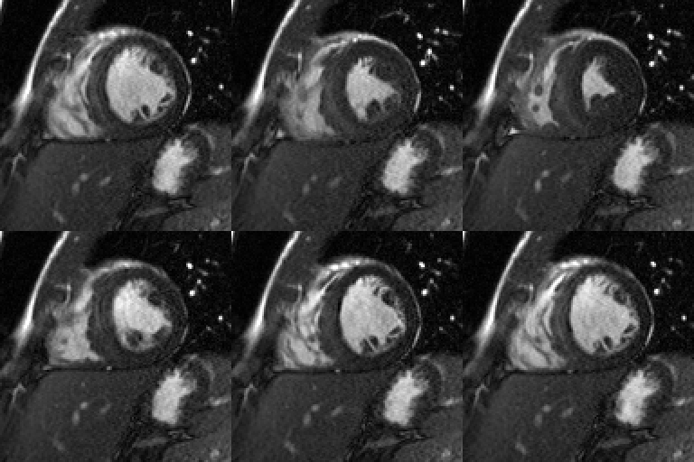
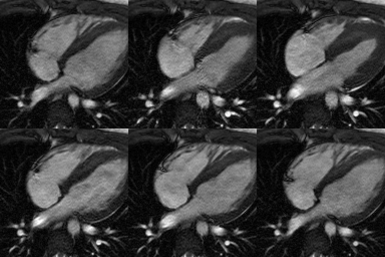
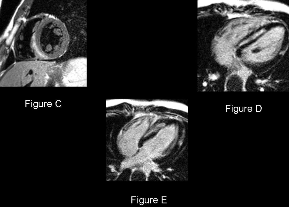
Selected Cardiac MRI images: Short axis SSFP (Fig. A) and 4-chamber SSFP (Fig. B) cine images demonstrate left ventricular hypertrophy, mild global hypokinesis, and a preserved ejection fraction. T2WI short axis view (Fig. C) reveals a mesocardial curvilinear band of high signal in the septal and inferoseptal wall of the left ventricle. 10 minute post-contrast myocardial signal–nulled T1-weighted 4-chamber views (Fig. D and E) show delayed mid-wall septal mesocardial and left ventricular free wall subepicardial hyperenhancement Diagnosis: Viral myocarditis (adenovirus)
Differential Diagnosis
Myocarditis of other etiologies
Background
Discussion
The myocardium is subdivided into subendocardial, subepicardial, and mesocardial regions. The subendocardium is the innermost region, lying in closest proximity to the ventricular blood pool, and is the region most sensitive to ischemia and infarction. The subepicardium is the outermost region, located farthest away from the blood pool and in close proximity to the pericardium. Subepicardial enhancement is seen in the majority of cases of myocarditis, however focal areas of transmural enhancement may be seen in severe cases. The mesocardium or midmyocardial region lies between the subendocardium and subepicardium. Myocarditis and various infiltrative cardiomyopathies can cause delayed enhancement in the midinterventricular septum. However, the abnormalities will not be limited to the midinterventricular septum, but will more pronounced in other myocardial regions as well. The distribution and pattern of delayed enhancement allows radiologist to differentiate ischemic from non-ischemic disease (Table -1) and thus approriately guide patient management. Patterns of abnormal delayed contrast enhancement include diffuse, patchy, and nodular.
Table-1: Differential Diagnosis based on Pattern of Delayed Contrast Enhancement
Subendocardial Vascular Subendocardial Non-vascular Subepicardial Mesocardial Transmural Ischemia Amyloidosis Myocarditis Hypertrophic Cardiomyopathy Infarction Infarction Loeffler Endocarditis Sarcoidosis Dilated Cardiomyopathy Myocarditis (severe) Histiocytoid Cardiomyopathy Pulmonary Artery Hypertension Sarcoidosis (chronic)
Clinical Findings
Myocarditis is a very common cause of nonischemic cardiomyopathy. The clinical presentation is often non-specific, ranging from vague to severe chest pain; to frank heart failure and sudden death. Markers of myocardial damage, tropinin or creatine kinase are elevated. Viral infection (parvovirus B-19; adenovirus) is the most common cause of myocarditis. Other possible causes include various drug toxicities and autoimmune disorders such as systemic lupus erythematosus (SLE) and mixed connective tissue diseases (MCTD). Because the clinical presentation is often nonspecific, diagnosis is dependent on clinical suspicion, laboratory data, and endomyocardial biopsy in select cases. When cardiac MR imaging findings are concordant with the clinical suspicion, biopsy may potentially be avoided.
Cardiac MRI Imaging Findings
- Delayed subepicardial contrast enhancement; frequently involves the lateral wall (Fig. D and E)
- Wall motion abnormalities in affetced regions (Fig. A and B)
- Increased T2 signal in affected regions (Fig. C)
- Ventricular dilatation
Prognosis
- Most patients: fully recover with supportive measures
- One-third of patients: develop dilated cardiomyopathy; may require cardiac transplantation
- An acute myocardial infarction-like syndrome with normal coronary arteries: good prognosis
- Heart failure, even with dilated left ventricle: may still have a good prognosis
- Ventricular dysrrhythmias and high-degree heart block: poor prognosis
- Loss of right ventricular function: strong predictor of death
CAVEATS
- Ischemic , or infarct-related, cardiomyopathy: tends to cause delayed enhancement in the subendocardium or transmurally and follows a specific vascular territory
- Nonischemic cardiomyopathies : tend to cause delayed enhancement in more than one vascular territory when restricted to the subendocardium or to spare the subendocardium
- Delayed subepicardial contrast enhancement: highly suggestive of myocarditis
- Myocarditis causes up to 20% of all cases of sudden death
- Delayed contrast enhancement limited to mesocardium in the midinterventricular septum: most often associated with hypertrophic cardiomyopathy (HCM) and dilated cardiomyopathy
Selected Readings
- Cummings KW, Bhalla S, Javidan-Nejad C, Bierhals AJ, Gutierrez FR, Woodard PK. A Pattern-based Approach to Assessment of Delayed Enhancement in Nonischemic Cardiomyopathy at MR Imaging. RadioGraphics 2009; 29: 89-103.
- Grizzard JD, Judd RM, Kim RJ. Teaching File Case 33. In: Cardiovascular MRI in Practice. Springer-Verlag, London; 2008: 130-131.
- Grizzard JD, Judd RM, Kim RJ. Teaching File Case 138. In: Cardiovascular MRI in Practice. Springer-Verlag, London; 2008: 130-131.
Original case written by its authors at Virginia Commonwealth University and published at this address as part of a weekly teaching collection. Reproduced here as an archive.