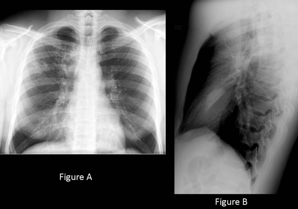
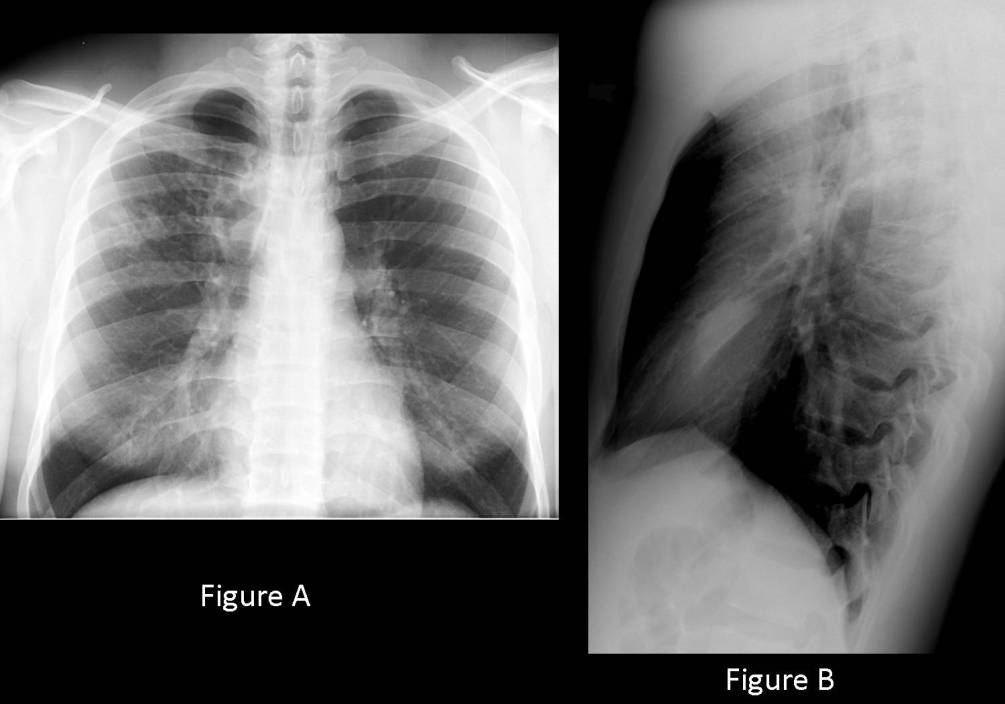
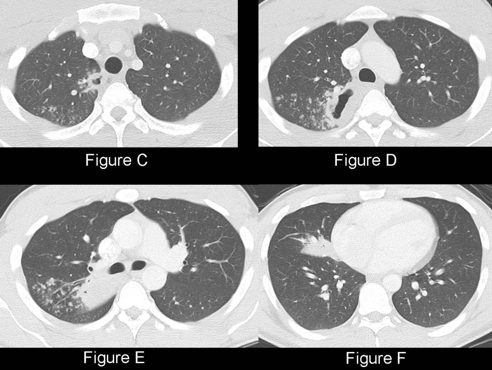
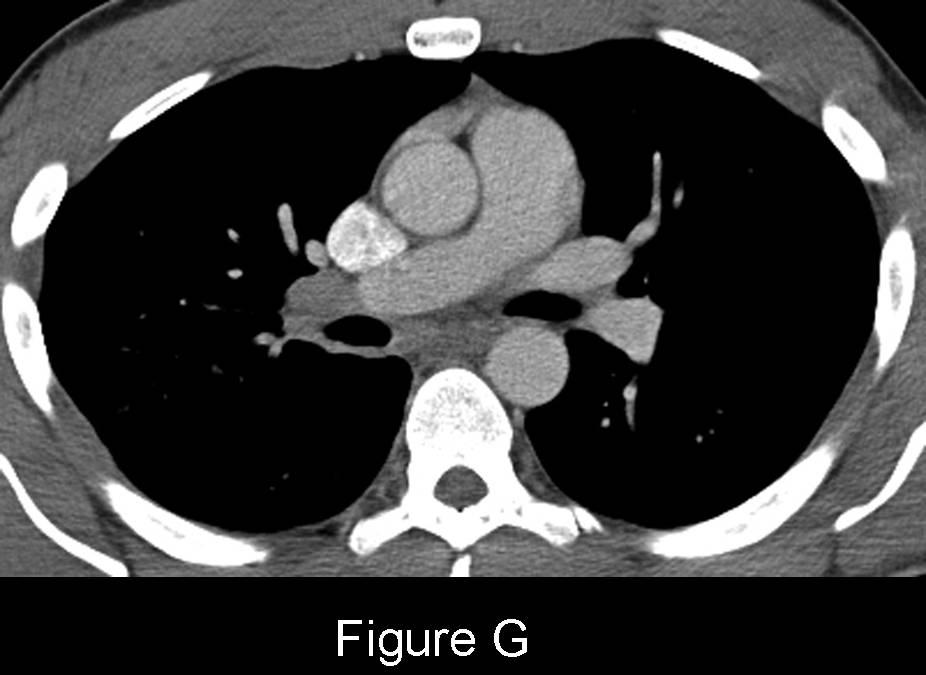
Initially withheld. Middle-aged man with productive cough, weight loss and night sweats. PA (Fig. A) and lateral (Fig. B) chest radiographs demonstrate a patchy focus of air space disease in the posterior segment right upper lobe with associated nodularity, eccentric cavitation and consolidation, and right hilar lymphadenopathy. Additionally, there is consolidation in the right middle lobe, best seen on the lateral exam (Fig. B). No pleural effusion is seen. Chest CT images (Fig. C and Fig. D) (lung windows) confirm two cavitary lesions in the right upper lobe, each with eccentric wall thickening. The first resides in the tracheoesophageal groove (Fig. C) and the second in the medial aspect of the posterior segment (Fig. D). Extensive tree-in-bud opacities are seen about these cavities and throughout the right upper lobe (Fig. C-E). Focal right middle lobe consolidation (Fig. F) (lung windows) and right hilar lymphadenopathy is also confirmed (Fig. G) (mediastinal windows) Diagnosis: Post-Primary Mycobacterium tuberculosis
Clinical Pearl: The radiographic findings are highly suspicious for active tuberculosis and warrant respiratory isolation and sputum cytology for acid fast bacilli until the diagnosis is otherwise excluded.
Differential Diagnosis
- Bacterial pneumonia with abscess formation
- Other infectious granulomatous diseases, including fungi and atypical organisms
- Primary and secondary lung neoplasia
Background
Discussion
Tuberculosis (TB) is a contagious disease, spread by the airborne transmission of Mycobacterium tuberculosis (MTB). MTB is one of the leading causes of infectious death in the world. Infected persons transmit bacilli via aerosolized droplets. The droplets enter the mid and lower lungs, where bacilli are phagocytized by alveolar macrophages. The initial immune response eventually leads to granuloma formations and parenchymal disease known as the Ghon focus. Inclusion of regional and hilar lymph nodes via lymphatic channels creates the Ranke complex. The initial infection is often clinically silent. 5-10% of those infected will develop active TB infection in their lifetimes, and at a much higher rate if co-infected with HIV-AIDS.
Approximately 1/3 of the world’s population is currently infected with MTB, with the largest number of new cases (2008) occurring in South-East Asia. In the United States, there were 12,904 cases of MTB reported in 2008 in the 50 states and District of Columbia, combined. This represents a 2.9% decrease from 2007, however, 18 states reported increased numbers of cases. California, Texas, New York, and Florida account for 49% of the national total new cases.
Individuals are greatest risk in the United States include those in ethnically diverse populations, immigrants, minorities, the homeless, the immunocompromised, the elderly, and the incarcerated.
Clinical Findings
Latent MTB refers to the development of conversion to a positive tuberculin skin test (purified protein derivative, PPD) in an asymptomatic person without active tuberculosis. Patients with active MTB have clinical, radiologic, and or laboratory signs of infection. Primary MTB refers to disease at the site of initial bacteria deposition and may manifest as severe cavitary pneumonia (progressive primary MTB). Post-primary MTB is caused by reactivation of dormant organisms or exogenous re-infection and typically involves areas of the lung with high oxygen tension (apical and posterior segments of upper lobe). Pulmonary symptoms may include: cough; hemoptysis; pleuritic chest pain; and rarely dyspnea. Constitutional signs include fever; weight loss; anorexia; weakness; and malaise. Patients with both primary and post-primary MTB may also be asymptomatic.
Imaging Findings
Chest Radiography
Primary MTB
- Consolidation
- Usually right-sided, dense, homogeneous; segmental, lobar, or multi-focal; may be associated with ipsilateral lymphadenopathy; cavitary consolidation in progressive MTB
- Lymphadenopathy
- Typically unilateral; usually right hilar and or paratracheal; more common in pediatric patients
- Atelectasis
- Usually lobar and right-sided; 30% of pediatric patients
- Pleural effusion
- Self-limited
- Unilateral or bilateral; more common in adults
- Ranke complex calcification
Post-Primary MTB
- Consolidation (Fig. A and Fig. B)
- Apical and posterior segments upper lobes (85%) (Fig. A and Fig. B)
- Superior segments lower lobes (14%)
- Ill-defined borders (Fig. A and Fig. B)
- Satellite nodules (Fig. A and Fig. B)
- Cavitation (45%) (Fig. A and Fig. B)
- Thin or thick wall
- Focal or multi-focal
- Air-fluid levels
- Perihilar nodular or linear opacities
- Atelectasis
- Multifocal ill-defined 5-10 mm air-space nodules (Fig. A and Fig. B)
- Tuberculoma
- Solitary or multiple pulmonary nodules
- Variable size
- Well-defined or ill-defined; may calcify
- Pleural disease
- Unilateral effusion
- Loculated effusion
- Pleural calcifications
- HIV-AIDS
- Lymphadenopathy +/-consolidations
- Lower frequency of cavitation
- Miliary nodules
CT
- Subtle cavitation (Fig. C-F)
- Centrilobular and tree-in-bud opacities (Fig. C-F)
- Ill-defined nodules (4-8 mm) (Fig. C-F)
- Lobular consolidations
- Thickened interlobular septa
- Peripheral rim-enhancement of lymph nodes
- Tuberculoma
- Nodule with rim enhancement
- Nodule with calcification
- Satellite lesions
- Bronchiectasis and or bronchostenosis
Complications
- Empyema; Empyema Necessitatis
- Pleural Calcification; Calcific Pleuritis
- Bronchostenosis; Bronchopleural Fistula
- End-stage Lung Disease
- Miliary Disease
- Mycetoma in Chronic Cavities
Management
- Clinical and radiographic suspicion warrants respiratory isolation
- First line drugs: isoniazid, rifampin, streptomycin, ethambutal, pyrazinamide
- Directly Observed Therapy (DOT)
- Increased prevalence of Multi-drug Resistant (MDR) TB, and now extensively drug resistant (XDR) TB
Selected Readings
- Reported Tuberculosis in the United States, 2008. Published 2009. Centers for Disease Control and Prevention. http://www.cdc.gov/tb/ World Health Organization Fact Sheet No. 104. Tuberculosis. March 2010.
- Muller NL, Silva CIS. Imaging of the Chest . Saunders, 2008. Pulmonary tuberculosis. 322-41.
- Parker MS, Rosado-de-Christianson ML, Abbott GF. Case 60: Mycobacterium tuberculosis pulmonary infection In:. Teaching Atlas of Chest Imaging . Thieme, New York. 2006. 243-48.
Original case written by its authors at Virginia Commonwealth University and published at this address as part of a weekly teaching collection. Reproduced here as an archive.