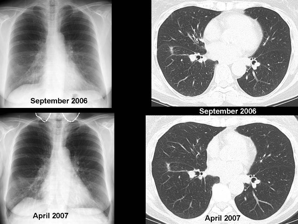
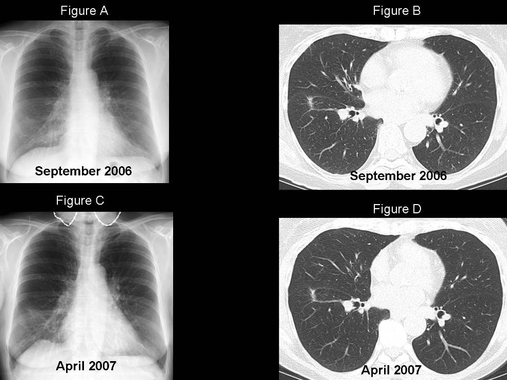
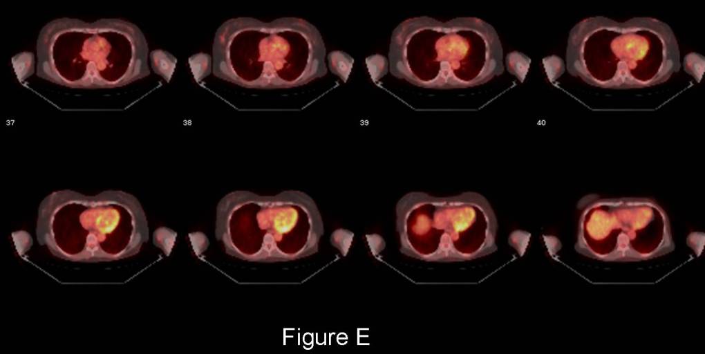
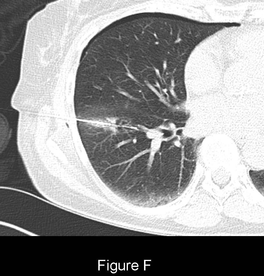
PA chest x-ray (September 2006) (Fig. A) with accompanying CT (September 2006) (Fig. B) (lung windows) shows a focal, fairly well-defined, spiculated 8 mm sub-solid nodule with surrounding ground-glass and desmoplasia of neighboring vessels in the superior segment of the right lower lobe. The patient was placed on antibiotics and a repeat study was performed to reassess the status of this lesion. The follow-up PA chest x-ray (April 2007) (Fig. C) and accompanying CT (April 2007) (Fig. D) (lung windows) show little if any change in appearance of this lesion. Subsequent FDG-PET scan revealed no appreciable metabolic activity in the lesion (Fig. E). A successful CT-guided percutaneous needle aspiration biopsy was then performed (Fig. F). Diagnosis: Bronchioloalveolar Carcinoma (BAC)
Differential Diagnosis
- Chronic Organizing Pneumonia
- Cryptogenic Organizing Pneumonia
- Scar
- Atypical Adenomatous Hyperplasia (AAH)
- Mixed subtype adenocarcinoma
- Metastasis
Background
Discussion
Bronchioloalveolar Carcinoma (BAC) is a subtype of adenocarcinoma with distinctive morphologic and prognostic features. It represents approximately 2-6% of all non-small cell lung cancers. BAC typically exhibits an indolent course with a pure lepidic growth pattern (i.e., grows along pre-existing structures without pleural, stromal or vascular invasion). Any evidence of invasive growth changes the diagnosis from pure BAC to adenocarcinoma, mixed subtype. There are three recognized subtypes of BAC: mucinous; nonmucinous; and mixed. The sub-solid nodule may represent one of the three peripheral types of adenocarcinomas: AAH; BAC; and mixed subtype adenocarcinoma. Alternatively, BAC may present with a multifocal distribution (25%), unlike other lung cancers (5%). BACs and BAC-related lesions exhibit prolonged doubling times relative to other primary lung neoplasms as illustrated in the table below:
Lesion Doubling Time Bronchioloalveolar Carcinoma (BAC) 567 days Atypical Adenomatous Hyperplasia (AAH) 988 days Mixed Subtype Adenocarcinomas 384 days
Etiology
BAC closely resembles AAH, the postulated precursor lesion of adenocarcinoma. Studies suggest that BAC is either closely associated with AAH or develops from AAH.
Clinical Findings
Approximately 50% of BACs are diagnosed in asymptomatic patients with incidentally discovered solitary pulmonary nodules. Patients with BAC that manifests with consolidation, or multifocal nodules, masses, and consolidations, may present with cough and fever. Mucin-producing BACs with extensive lung involvement may result in bronchorrhea. The latter may be complicated by hypovolemia and electrolyte imbalance.
Imaging Findings
Chest Radiography
- Variable-sized peripheral solitary pulmonary nodule or mass (Fig. A; Fig C)
- Less frequently, focal consolidation, multifocal nodules, masses and/or consolidation(s)
MDCT
- Purely ground-glass opacity (GGO) or a GGO with solid components (sub-solid nodule) (Fig. B; Fig. D)
- Heterogeneous attenuation:
- Ground-glass opacity
- Air bronchograms
- Air bronchiolograms
- Intratumoral “bubble-like” cystic airspaces
- Irregular or ill-defined margins, pleural tails (Fig. B; Fig. D)
- Multifocal ground-glass opacities, consolidations and/or nodules
- CT angiogram sign; in setting of consolidation
- CT bronchus sign; in setting of consolidation
- Insidious growth with long doubling times
Management
Lesion Follow-up / Management Isolated GGO lesions:
GGO < 5 mm No Follow-up GGO 5-10 mm Follow-up in 3-6 months If persists, annual follow-up x 3 years GGO > 10 mm Follow-up in 3-6 months If persists, surgical resection Any mixed isolated sub-solid lesion; regardless of size FDG-PET; Biopsy; Surgical Resection
Multiple lesions
Pure GGO > 5 mm F/U in 1 year Pure GGO 5–10 mm F/U in 1 year GGO or mixed with dominant lesion >10 mm Surgical resection
Prognosis
- Prognosis is based on the Noguchi type classification
Noguchi Type Classification 5-Year Survival rate
Type A (Localized BAC)
Type B (Localized BAC with foci structural collapse)
100% Type C (Localized BAC; active fibroblastic proliferation) 75% Type D – F (Poorly differentiated tubular, papillary adenocarcinoma) 50%
Caveats
- While other primary lung cancers may also present as GGOs, metastatic lesions rarely do so. This finding may help narrow the differential diagnosis
- Unfortunately, do to the high false-negative rate with slow growing tumors like BAC and carcinoid tumors, FDG-PET is often of little value
- Diagnostic yield of CT-guided FNA and CT-guided core needle biopsy is only 51% and 73%, respectively. Thus, obtaining a definitive diagnosis prior to treatment can be problematic
Selected Readings
- Godoy MCB, Naidich DP. Subsolid Pulmonary Nodules and the Spectrum of Peripheral Adenocarcinomas of the Lung: Recommended Interim Guidelines for Assessment and Management. AJR 2009; 253: 3; 606-622.
- Parker MS, Rosado de-Christenson ML, Abbott GF. Case 73 Lung Cancer: Bronchioloalveolar Carcinoma. In: Teaching Atlas of Chest Imaging 2006; Thieme, New York: 296-299.
Original case written by its authors at Virginia Commonwealth University and published at this address as part of a weekly teaching collection. Reproduced here as an archive.