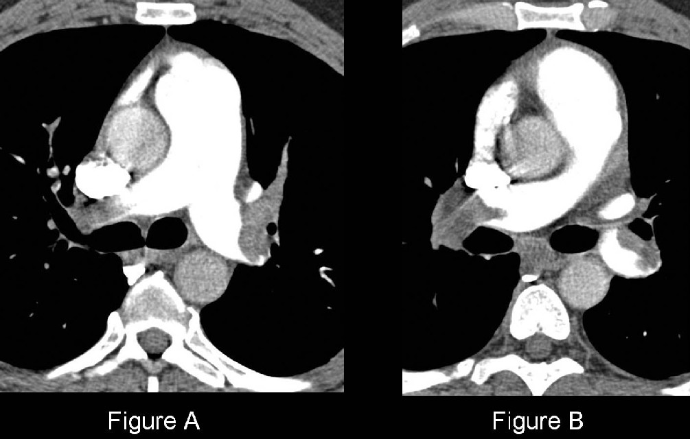
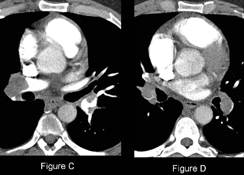
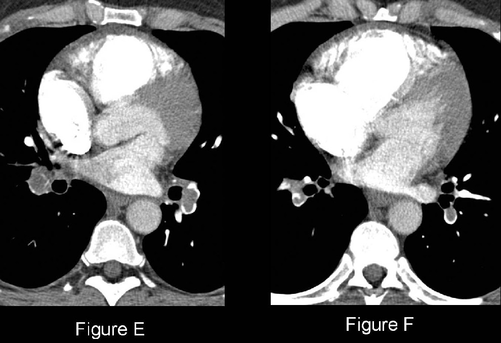
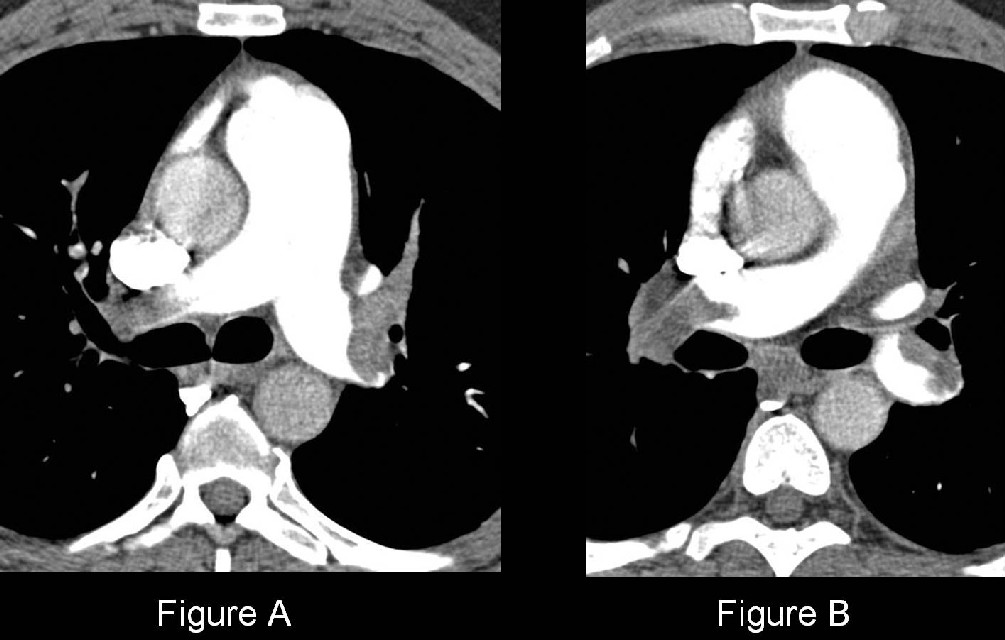
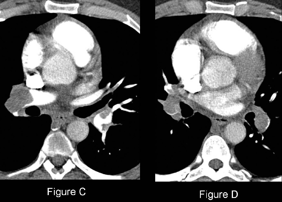
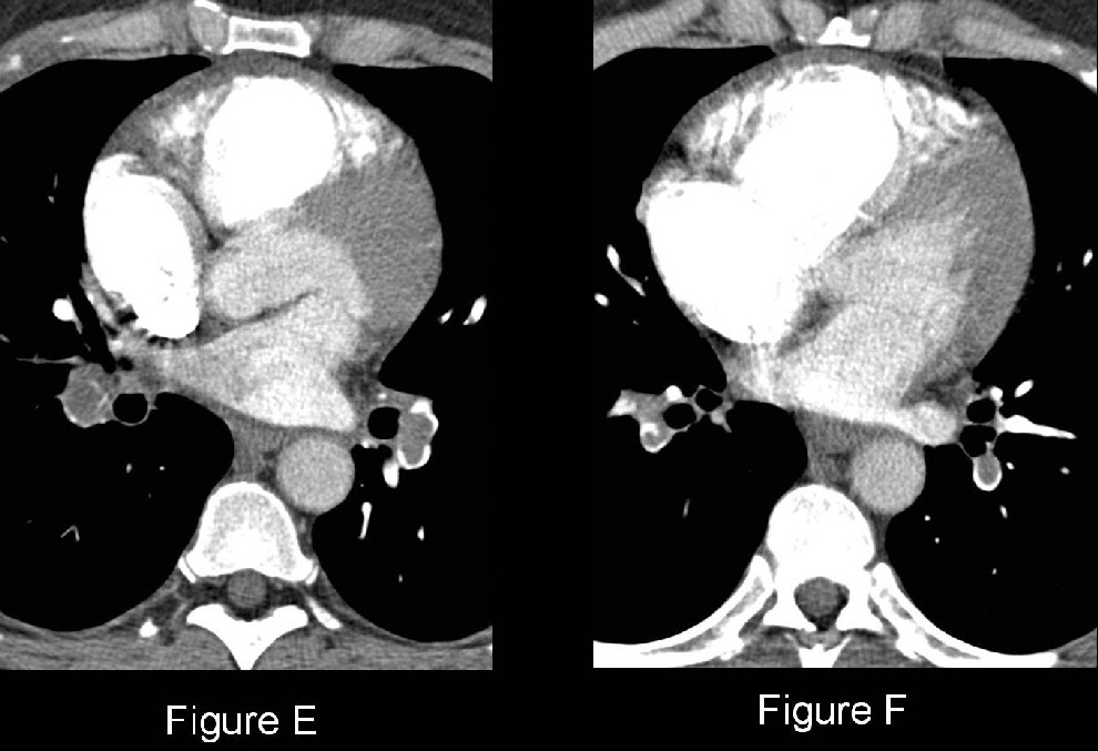
Chest CTPA (mediastinal windows) (Fig. A-F) demonstrates extensive soft-tissue filling defects involving virtually every branch of the pulmonary arterial circuit. The right heart and right ventricle in particular is enlarged and there is inward bowing of the interventricular septum. Diagnosis: Acute Pulmonary Thromboembolic Disease; Right Heart Strain
Differential Diagnosis
None
Background
Discussion
Pulmonary thromboembolism (PTE) is the third most common cause of cardiovascular death, after myocardial ischemia and stroke. However, PTE is not a disease in and of itself but rather a complication of underlying venous thrombosis. Venous thromboembolism (VTE) continues to be a significant health problem. The average annual incidence of VTE is 1 per 1,000 (U.S.A.) with about 250,000 incident cases occurring annually. Autopsy studies show an additional equal number of patients with PTE-VTE unsuspected or not diagnosed antemortem. Thus, it is estimated that 650,000 to 900,000 cases of fatal and nonfatal PTE-VTE occur annually in the United States alone. Acute PTE is responsible for 2-7% of acute care hospital deaths.
Etiology
Normally, there is a dynamic equilibrium in the venous system between the formation and subsequent lysis of microthrombi which allows for local hemostasis in response to injury but prevents uncontrolled propagation of thrombus. Certain pathological conditions allow microthrombi to escape the fibrinolytic system, propagate, and potentially embolize distally into the pulmonary arterial circuit as pulmonary emboli. In fact, 90% of such pulmonary emboli originate from lower extremity deep veins. Predisposing VTE is induced by inflammation of the vessel wall, venous stasis, and hypercoagulable states (i.e., Virchow triad). Clinical risk factors for PTE-VTE are directly related to one or more of these components and are summarized in Table 128-1.
Table -1. Clinical Risk Factors for Developing PTE-VTE
Hereditary Acquired Prolonged Immobilization Acute Illness Neoplasia Pregnancy Other Protein C Deficiency Trauma ICU Chemotherapy Peri-partum OCP * Protein S Deficiency Recent Surgery Heart Failure
Post-partum Long-term Catheters Anti-thrombin III Deficiency Burns AIDS (lupus anticoagulant)
Pacemakers Factor V Leiden Fractures Acute MI
Advanced Age Plasminogen or Plasminogen Activator Abnormalities Obesity New-onset Atrial Fibrillation
Previous PTE-VTE Fibrinogen Abnormalities Spinal Cord Injuries Polycythemia
High-dose Estrogen Therapy
Travel SLE
Varicose Veins OCP *-oral contraceptives
Clinical Findings
The diagnosis of PTE poses a challenge for both clinicians and radiologists because the signs and symptoms are nonspecific. The classic triad of pleuritic chest pain, dyspnea, and hemoptysis likewise is neither sensitive nor specific and occurs in less than 20% of patients diagnosed with PTE. Additionally, many patients are relatively asymptomatic. Physical signs of PTE may include: tachypnea; rales, pleural rub or new wheezing; tachycardia; S3 or S4 gallop; new murmur; accentuated second heart sound; fever; diaphoresis; lower extremity edema; and cyanosis. Massive pulmonary embolism may be associated with hypotension due to acute cor pulmonale. Patients with PTE may have an abnormally high A-a gradient, and right ventricular strain on ECG. D-dimer levels may be elevated with PTE-VTE. D-dimer is a degradation product of plasmin-mediated proteolysis of cross-linked fibrin and is considered positive when > 500 ng/mL. However, levels may be falsely elevated in numerous clinical settings (e.g., severe trauma, recent surgery, infection, neoplasia, rheumatoid factor, and advanced age). A negative D-dimer is most valid and useful when done on patients considered low-risk for PTE-VTE or when combined with other data (e.g., Wells criteria).
Imaging Findings
MDCT Pulmonary Angiography (CTPA)
New gold standard for ruling-in or ruling-out diagnosis of PTE
Diagnostic criteria (direct signs)
- Complete arterial occlusion with failure to opacify the entire lumen; artery may be enlarged in comparison with pulmonary arteries of the same order of branching (Fig. A-F)
- Central arterial filling defect surrounded by IV contrast material (Fig. A-F)
- Peripheral intraluminal filling defect that makes an acute angle with the arterial wall (Fig. D-F)
Indirect Signs
- Atelectasis; often subsegmental
- Small pleural effusion
- Oligemia of affected segment
- Pulmonary hemorrhage or infarct
- Right heart strain (Fig. F)
- Right ventricular dilatation (right ventricular cavity wider than left ventricular cavity in short axis); +/- reflux of contrast media into hepatic veins
- Deviation of interventricular septum towards the left ventricle
Potential CT pitfalls
- Confusion of filling defects with hilar and infrahilar lymph nodes
- Poor vessel opacification and motion artifact mimicking or obscuring filling defects
- Increased noise (i.e., quantum mottle) in obese patients
- Obscuration of vessels by adjacent regions of parenchymal consolidation
- Segmental or subsegmental bronchial mucus plugs
Treatment
- All patients require anticoagulation
- Thrombolysis and/or thrombectomy may be indicated in select patients (i.e., extensive PTE; concomitant moderate to severe right ventricular dysfunction despite preserved systemic arterial pressure)
- IVC filters may have to be deployed in patients with contraindications to anticoagulation therapy
Prognosis
- 1/3 of patients with PTE will have recurrent emboli
- Mortality massive PTE (i.e., systolic arterial pressure < 90 mm Hg; 4% patients): between 30% and 60%; majority of deaths occur in the first 1-2 hours
- Mortality for patients with non-massive PTE (i.e., systolic arterial pressure ≥ 90 mm Hg; 96% patients): < 5% in the first 3-6 months of anticoagulation therapy
- Shock Index (Heart Rate ÷ Systolic Blood Pressure) >1 has been used as a predictor of in-hospital deterioration of patients diagnosed with acute PTE
- Troponin levels are commonly elevated in acute PTE, especially in patients with significant clot burden and right heart dysfunction; associated with worse short-term and long-term prognosis and up to 5X increase in mortality
Caveats
- Most important clinically identifiable risk factor for PTE-VTE; prior history of such
- Considerable advantage of CTPA over both V/Q scan and catheter pulmonary angiography is its ability to depict other conditions that clinically mimic PTE (e.g., aortic dissection, pneumothorax, pleural or pericardial disease, pneumonia, lung abscess, pneumomediastinum, esophageal rupture, mediastinitis, pulmonary fibrosis, and neoplastic disease). Such alternative diagnoses have been reported in 11-70% of CTPA examinations performed for clinically suspected acute PTE
- Paradoxical systemic arterial embolism (PSAE) is a rare complication of PTE (2%). PSAE requires passage of venous thrombus into the arterial circulatory system through a right-to-left shunt, most commonly a patent foramen ovale (PFO). Complications include stroke, myocardial infarction, and showering of thrombi to kidneys, superior mesenteric artery, and lower extremities. PFO may be confirmed by an echosonographic bubble study.
Selected Readings
- Grifoni S, Olivotto I, Cecchini P, et al.: Short-term clinical outcome of patients with acute pulmonary embolism, normal blood pressure, and echocardiographic right ventricular dysfunction. Circulation 2000, 101:2817-2822.
- Nazaroglu H, Ozmen CA, Akay HO, Kilinc I, Bilici A. 64-MDCT pulmonary angiography and CT venography in the diagnosis of thromboembolic disease. AJR 2009; 192:654-661.
- Sandler DA, Martin JF. Autopsy proven pulmonary embolism in hospital patients: are we detecting enough deep vein thrombosis? J R Soc Med.1989; 82(4):203-205.
- Smita Patel and Ella A. Kazerooni. Helical CT for the Evaluation of Acute Pulmonary Embolism. AJR 2005; 185:135-149.
- Tapson VF. Acute pulmonary embolism. N Engl J Med. 2008; 358(10):1037-1052.
Original case written by its authors at Virginia Commonwealth University and published at this address as part of a weekly teaching collection. Reproduced here as an archive.