Differential Diagnosis
Various nerve sheath tumors
Lymphadenopathy
Discussion
Background
Extramedullary hematopoiesis (EMH) is the formation of blood elements, usually as a compensatory response to severe chronic anemia, outside the normal marrow space. This most commonly takes place in the liver, spleen, lymph nodes, and the posterior mediastinum (i.e., paravertebral space).
Etiology
EMH typically occurs in response to ineffective erythrocyte production or increased erythrocyte destruction (e.g., hemolysis). It is associated with severe congenital hemolytic anemias (e.g., thalassemia major, hereditary spherocytosis), polycythemia vera, lymphoproliferative disorders (e.g., myelofibrosis, chronic leukemia, lymphoma), and sickle cell disease. It has been proposed that mediastinal EMH results from paravertebral osseous extrusion of marrow elements through the thinned cortices of neighboring ribs and the vertebral column.
Clinical Findings
Mediastinal EMH is usually asymptomatic. Affected patients are typically diagnosed on chest radiography or other imaging studies acquired for unrelated reasons. Rarely mediastinal EMH may be complicated by spinal cord compression and spontaneous hemothorax.
Imaging Findings
Chest Radiography
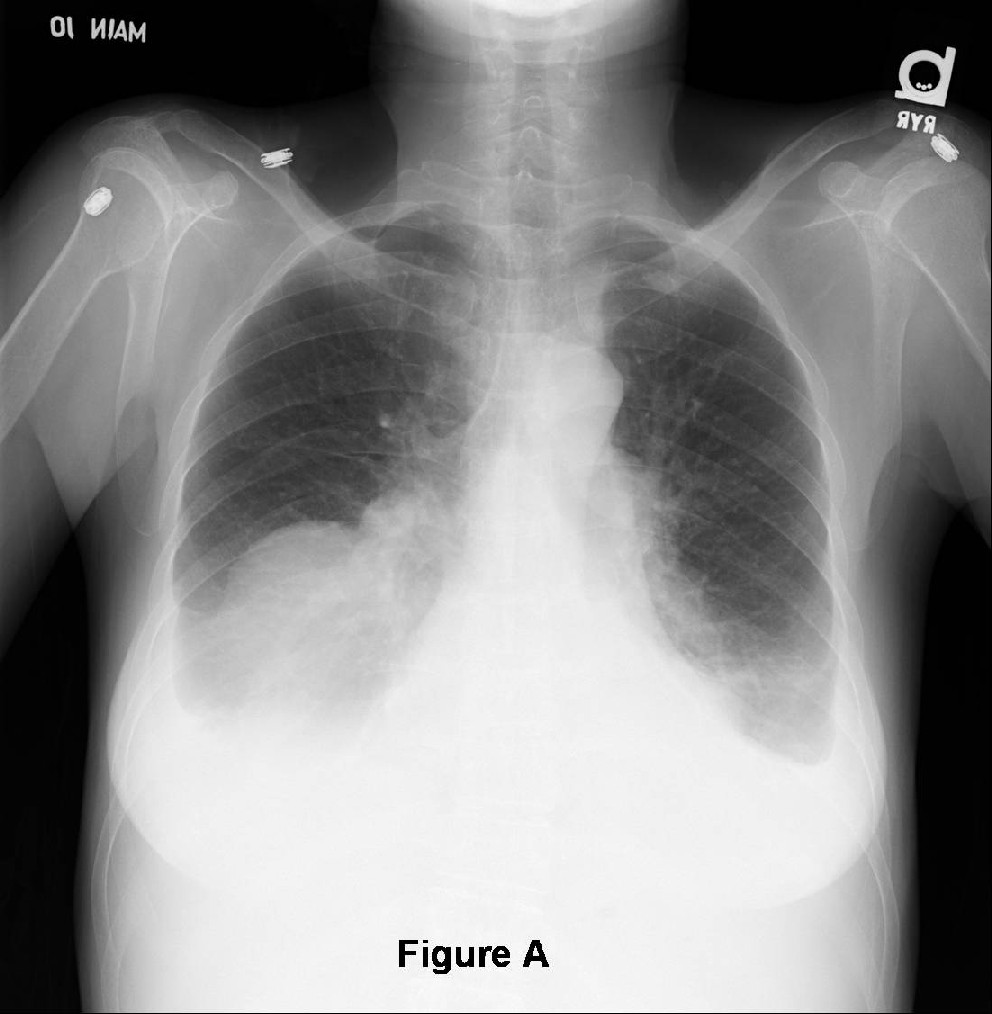
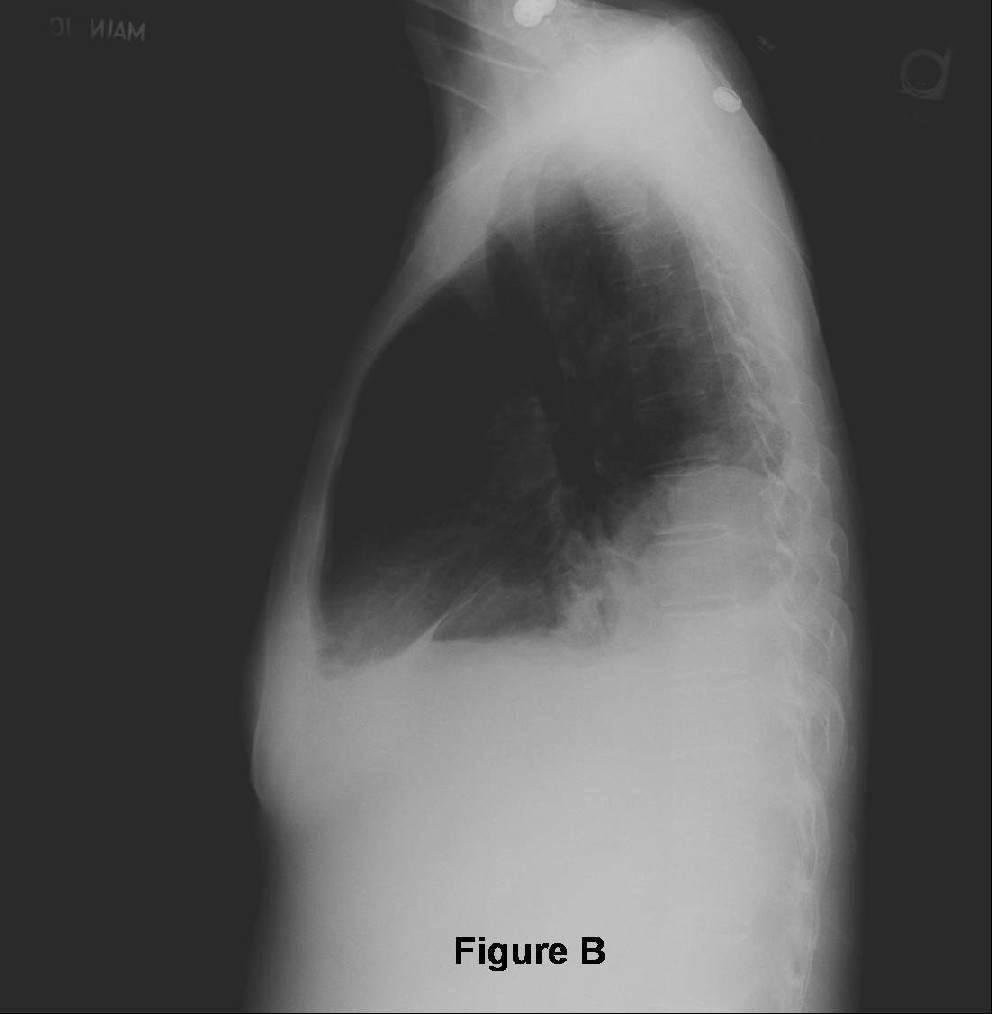
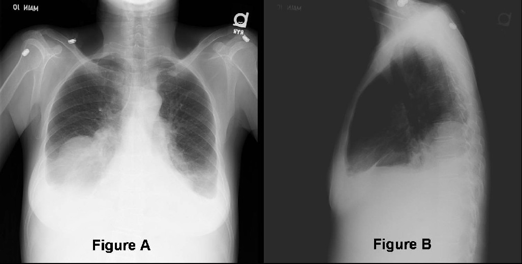
- Unilateral or bilateral, smooth, lobulated, posterior mediastinal-paravertebral mass(es) (Fig. A and Fig. B)
- Usually located between the 6th and 12th vertebral body in the inferior thorax (Fig. A and Fig. B)
- May span entire length of the paravertebral compartment (Fig. A and Fig. B)
- Ribs and thoracic spine may demonstrate “coarsened” trabeculae from expansion of the osseous erythroid bone marrow spaces
CT
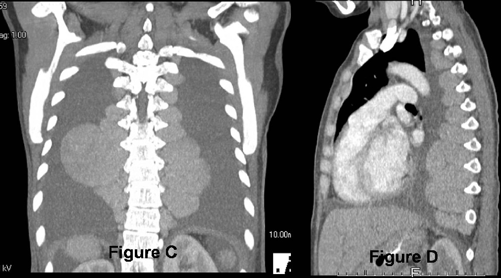
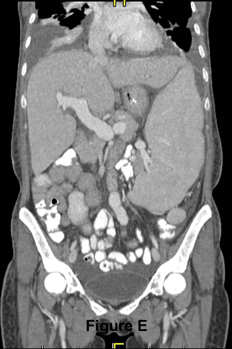
- Well-defined, lobulated, homogeneous paravertebral soft tissues masses (Fig. C-Fig. E)
- May contain fatty attenuation internal elements, especially in the setting of splenectomy
- Adjacent osseous structures may appear “lacy” with “coarsened features” secondary to marrow expansion.
- Rarely associated with an interstitial parenchymal pattern; ground-glass opacities; pleural effusion (Fig. C-Fig. D)
MR
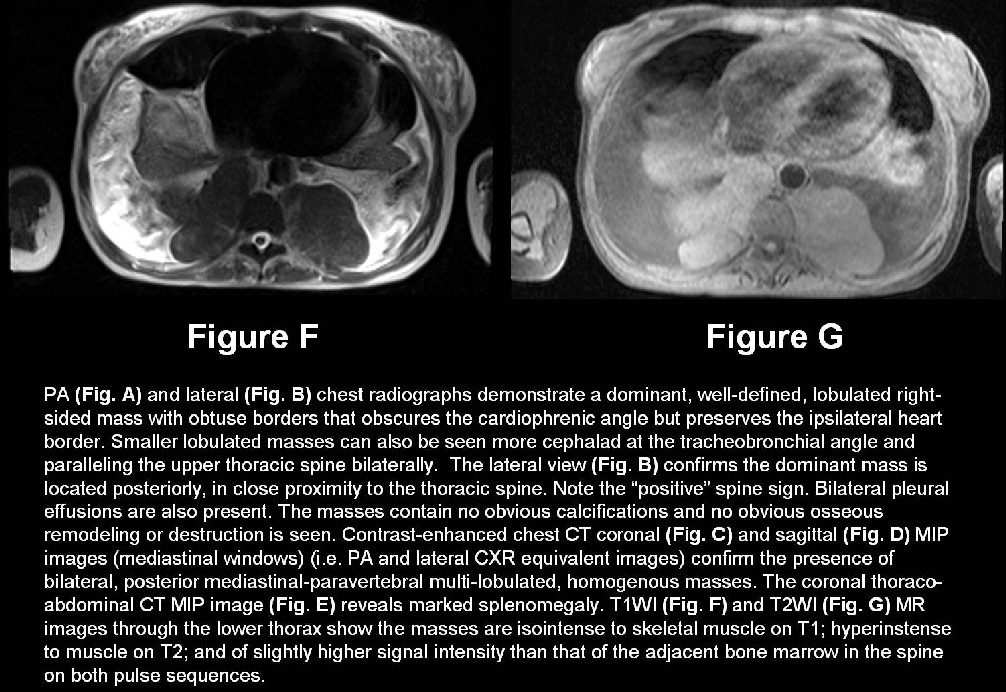
- T1WI- isointense to skeletal muscle (Fig. F and Fig. G)
- T2WI-hyperintense to skeletal muscle (Fig. F and Fig. G)
- Typically higher signal intensity than adjacent marrow on T1 and T2 (Fig. F and Fig. G)
- Intermediate enhancement of the masses after administration of a paramagnetic agent
- Vertebral bodies often have low-to-intermediate signal intensity as a result of displacement of fatty marrow by hematopoietic marrow
Nuclear Medicine
- Uptake of Tc-99m-labeled monoclonal antibodies
- Binds to hematopoietic cells
- Highly sensitive
- Tc-99m sulfur colloid
- Intense scintigraphic activity in the paraspinal masses
Management
- Unnecessary in asymptomatic patients
- Blood transfusions when necessary for symptomatic anemia
- Surgical decompression and/or radiation therapy for epidural involvement, spinal cord compression
Prognosis
- Variable and dependent upon the underlying predisposing condition
Selected Readings
- Gogia P, Goel R, Nayar S. Extramedullary paraspinal hematopoiesis in hereditary spherocytsosis. Ann Thorac Med 2008; 3(2): 64-66
- Lall C, Payne DK. A patient with anemia and a paraspinal chest mass. Chest 2003; 124(2): 732-734.
- Parker MS, Rosado-de-Christenson ML, Abbott GF. Case 179: Extramedullary hematopoiesis. In: Teaching Atlas of Chest Imaging, New York: Thieme, 2006: 695-698
- Tsitouridis J, Stamos S, Hassapopoulou E, Tsitouridis K Nikolopoulos P. Extramedullary paraspinal hematopoiesis in thalassemia: CT and MRI evaluation. Eur J Radiol 199; 30(1): 33-38.
Original case written by its authors at Virginia Commonwealth University and published at this address as part of a weekly teaching collection. Reproduced here as an archive.