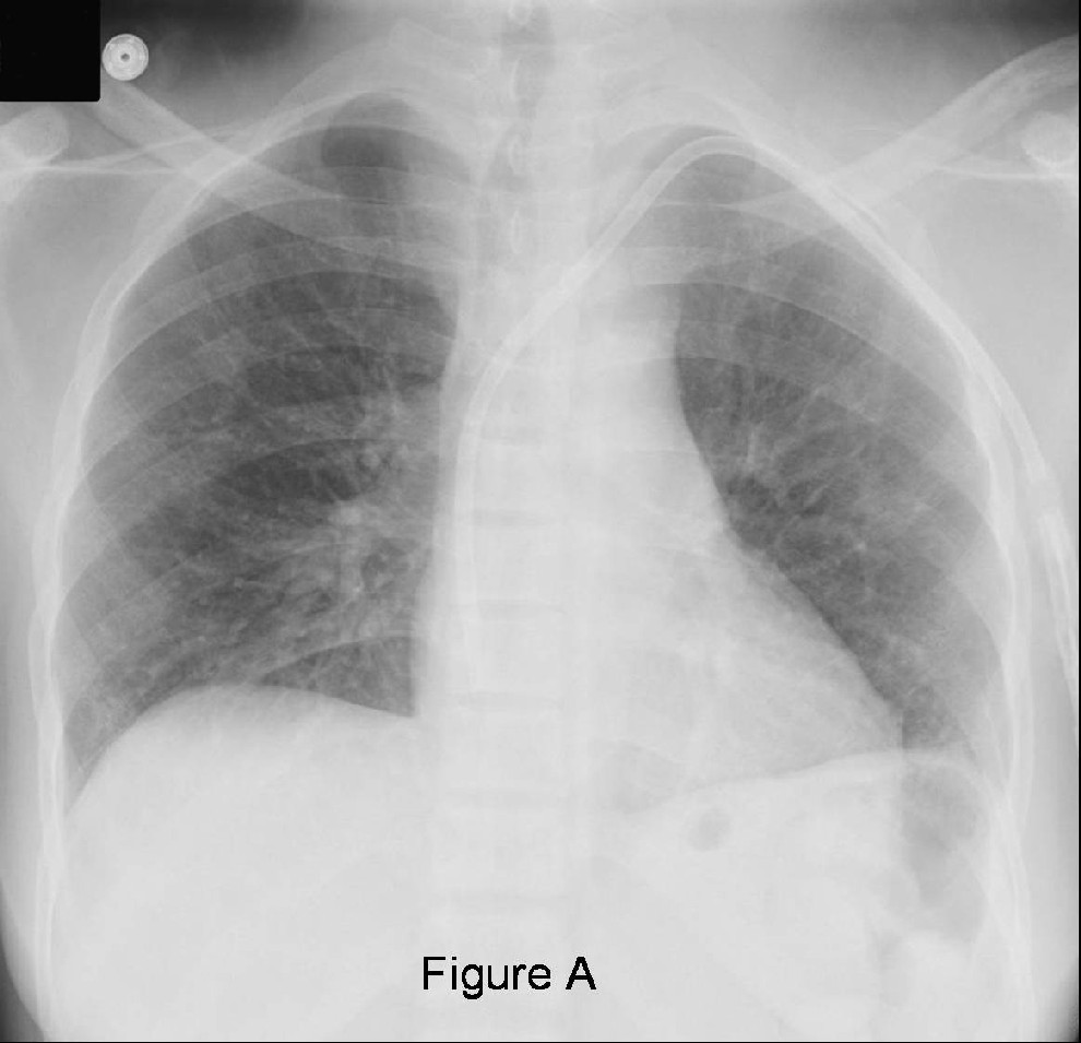
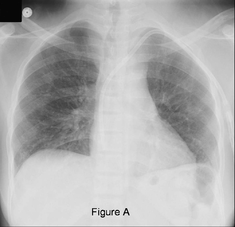
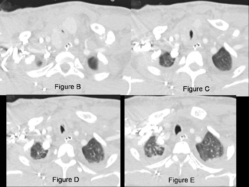
AP chest radiograph (Fig. A) demonstrates focal, short segment, symmetric narrowing of the proximal tracheal air column (arrows). Contrast-enhanced chest CT (Fig. B-E) (lung windows) confirms the presence of marked tracheal narrowing with a reduced coronal diameter and anterior luminal tapering. Diagnosis: Tracheal stenosis; post-intubation (tracheostomy) injury
Differential Diagnosis of Tracheal Stenosis
Congenital Infectious Inflammatory Neoplastic Traumatic Collagen Vascular Posterior fusion of tracheal rings Tuberculosis Amyloidosis Primary benign and malignant lesions Post-intubation Wegener granulomatosis Vascular rings Histoplasmosis Sarcoidosis Direct invasion Tracheotomy Tracheopathia osteoplastica Anomalous subclavian artery Blastomycosis Relapsing polychondritis Metastatic disease Blunt or penetrating Pulmonary sling Rhinoscleroma Fibrosing mediastinitis Thyroid goiter with extrinsic compression Inhalational burns Double aortic arch Crohn disease Radiation therapy Behçet syndrome Surgery Post-transplantation stenosis
Discussion
Background
The cross-sectional area of the trachea can be evaluated from PA and lateral chest radiography as well as CT. As measured 2.0 cm above the top of the transverse aorta, the normal dimensional range of the tracheal air column in men and women is as follows:
Patient Sex Coronal Diameter (mm) Sagittal Diameter (mm) Men 13-25 13-27 Women 10-21 10-23
Tracheal stenosis is defined as narrowing of the tracheal lumen by more than 10% of its normal diameter. It is a relatively uncommon condition with a frequently insidious onset. Early signs and symptoms may be disregarded or confused with other clinical disorders.
Etiology
The most common cause of both laryngeal and tracheal stenosis is trauma. Please see the above Table in the Differential Diagnosis Section. Of the potential list of trauma-related causes, prolonged endotracheal intubation is the leading cause of stenosis, primarily complicating multiple trauma victims or patients undergoing cardiovascular surgery. Once the ET tube or tracheostomy device is in position, the balloon cuff is inflated with enough air to occlude the tracheal lumen and create an airtight system. The airtight seal is formed by expanding the tracheal lumen and deforming the tracheal wall which subsequently compresses the tracheal mucosa. If the cuff pressure exceeds the mucosal capillary pressure (i.e.,> 20 mm Hg), ischemic necrosis ensues. Strictures may occur as early as 36 hours post-intubation. Utilization of ET tubes with low-pressure cuffs has reduced the prevalence of tracheal stenosis to < 1% as compared to a prevalence of 20% when high-pressure cuffs were more commonly in use.
Tracheal stenosis following the prolonged use of cuffed tracheostomy or ET tubes typically occurs in one of three locations: (1) stoma site; (2) level of the inflatable cuff itself; and (3) where the device tip impinged upon the tracheal mucosa.
Clinical Findings
Most patients are symptom free for a variable time frame following extubation or tracheostomy removal. Over time, affected patients may experience difficulty clearing secretion and go on to develop dyspnea on exertion, stridor and wheezing. Stridor typically develops five weeks or more after extubation.
Pathology
- Ischemic necrosis progresses to a superficial tracheitis and then to shallow muscosal ulcerations.
- Progressive mucosal erosion exposes the cartilaginous rings which soften and fragment
- Fibrosis subsequently occurs in the damaged tracheal wall leading to stenosis
Imaging Findings
Chest Radiography
- Post-intubation tracheal stenosis extends for several centimeters; typically affects the tracheal lumen above the thoracic inlet
- Tracheostomy-related stenosis is more cephalad; situated 1.5-2.0 cm or more distal to the stoma or in the vicinity of the stoma; extends 1.5-2.5 cm in length (2-4 cartilaginous rings) (Fig. A)
- Circumferential tracheal narrowing over a length of approximately 2.0 cm (Fig. A)
- Thin membrane or web of granulation tissue projecting at right angles from the tracheal wall
- Long, thickened, eccentric soft-tissue opacity compromising tracheal lumen
CT
- Circumferential or eccentric tracheal narrowing (Figs. B-E)
- Variable tracheal wall thickness (Figs. B-E)
- Degree and extent of tracheal narrowing is often underestimated on conventional radiography and axial CT images.
- Thin collimated images (1-3-mm) and multiplanar reconstructions, volume rendered and 3-D images, and virtual bronchoscopy may be necessary to fully appreciate the degree and extent of stenosis
Management
- Surgical excision of stenotic segment and reconstruction
- Endoscopic mechanical dilatation
- Tracheal stenting
- Laser photoablation for focal mucosal lesions
Prognosis
- Excellent; sleeve resection curative in 91% of patients
Caveats
- Tracheal stenosis as a complication of intubation or tracheostomy is one of the most common causes of chronic upper airway obstruction
- Tracheomalacia results from an abnormal degree of compliance of the tracheal wall and its supporting cartilage. The resultant flaccidity is usually apparent during forced expiration
- Tracheomalacia and / or ulcerative tracheoesophageal fistula may occur as sequelae of endotracheal intubation
Selected Readings
- Fraser RS, Müller NL, Coleman N, Paré PD. Upper Airway Obstruction. In: Fraser RS, Müller NL, Coleman N, Paré PD, eds. Fraser and Pare’s Diagnosis of Disease of the Chest, 4th ed. Philadelphia: Saunders, 1999: 2033-2035.
- Lee KS, Yoon JH, Kim TK, Kim JS, Chung MP, Kwon OJ. Evaluation of tracheobronchial disease with helical CT with multiplanar and three dimensional reconstruction: correlation with bronchoscopy. RadioGraphics 1997; 17:555-567.
- Parker MS, Rosado-de-Christenson ML, Abbott GF. Tracheal Stricture. In: Teaching Atlas of Chest Imaging. New York: Thieme, 2006: 103-106.
Original case written by its authors at Virginia Commonwealth University and published at this address as part of a weekly teaching collection. Reproduced here as an archive.