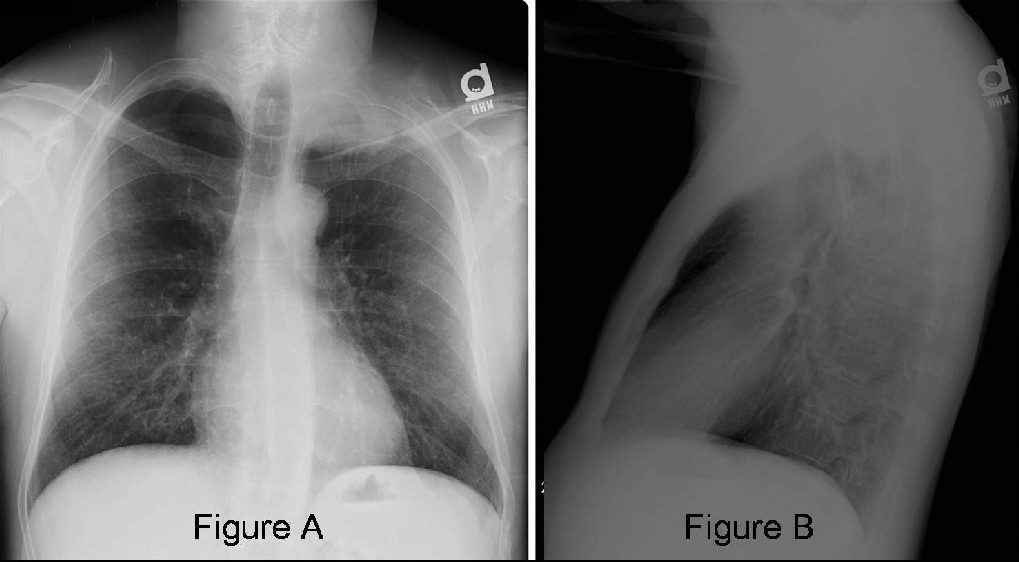
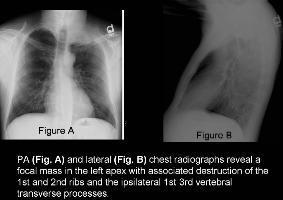
PA (Fig. A) and lateral (Fig. B) chest radiographs reveal a focal mass in the left apex with associated destruction of the 1st and 2nd ribs and the ipsilateral 1st-3rd vertebral transverse processes. Diagnosis: Pancoast tumor (also known as superior sulcus tumor)
Differential Diagnosis
· Neoplasia
o Primary Lung Cancer
· Usually non-small cell lung cancer NSCLC; rarely small cell lung cancer SCLC
o Other Primary Thoracic Neoplasms
· Sarcoma
· Mesothelioma
· Adenoid cystic carcinoma
· Hemangiopericytoma
o Metastatic disease
· Laryngeal cancer
· Thyroid cancer
· Uterine cervical cancer
· Urinary bladder cancer
o Hematologic Malignancies
· Lymphoma
· Plasmacytoma
· Lymphomatoid granulomatosis
o Nerve Sheath Tumor
· Neurofibroma
· Schwannoma
• Infection
o Bacterial
· Staphylococcal sp.
· Pseudomonas sp.
· Actinomycosis
o Fungal
· Aspergillosis sp
· Cryptococcus sp
o Mycobacterium tuberculosis
o Parasitic
· Miscellaneous
o Asymmetric apical cap
o Asymmetric apical extrapleural fat deposition
o Apical extension of or located pleural effusion
o Cervical rib syndrome
o Radiation fibrosis (e.g., antecedent head and neck cancer)
o Subclavian artery aneurysm
o Pulmonary amyloidoma
Discussion
Background
Primary bronchogenic or lung carcinoma may be referred to as a Pancoast tumor (or superior sulcus tumor, although the latter term lacks an anatomical basis) if it arises from the apex of the lung, the bulk of the tumor is extrathoracic, and the tumor principally invades the apical chest wall and or its associated neurovascular structures rather than the underlying lung parenchyma. In 1932, Dr. Henry Pancoast first described the radiographic findings associated with this primary lung tumor. These tumors are infrequent, comprising <5% of all bronchogenic carcinomas. Affected patients often present with a constellation of characteristic clinical signs and symptoms, collectively known as Pancoast Syndrome, which includes shoulder and arm pain, Horner’s syndrome with concomitant involvement of the sympathetic ganglia (i.e., miosis, ptosis, and ipsilateral facial anhydrosis), and weakness and atrophy of the upper extremity musculature, typically in the ulnar nerve distribution.
Etiology
Non small cell lung carcinoma (NSCLC) is the most common cell type of Pancoast tumor. Squamous cell is usually the most frequent cell type, followed closely by adenocarcinoma, and less frequently, large cell carcinoma. Rarely, small cell carcinoma (SCLC) (1-3% of cases) may be responsible. Various other pathologic processes may present as Pancoast tumors, such as metastatic solid tumors, hematologic malignancies, infections, cervical ribs, and pulmonary amyloidosis.
Clinical Findings
Most frequently, affected patients present with shoulder pain produced by invasion of the brachial plexus, parietal pleura, surrounding fascia, distal cervical or proximal thoracic spine, and adjacent ribs. Pain, weakness, and muscle atrophy often occur along the distribution of the eighth cervical nerve trunk and first and second thoracic nerve trunks, particularly in the ulnar nerve distribution. Horner’s syndrome, seen in up to 83% of affected patients, is caused by invasion of the paravertertebral sympathetic chain and the inferior cervical ganglion. Interestingly, even patients without frank brachial plexus invasion by tumor may present with a brachial plexopathy which is often relieved following surgical resection or tumor debulking. Infrequently, patients with a Pancoast tumor may present with a paraneoplastic syndrome. Most often these syndromes are the result of secretion of various hormones or metabolites by the tumor itself. Some of the more common paraneoplastic syndromes include Cushing syndrome, increased antidiuretic hormone (ADH) secretion, hypercalcemia, various myopathies, hematologic disorders, and hypertrophic osteoarthropathy (HPOA). Although the presence of paraneoplastic syndromes does not indicate tumor unresectability, most of these syndromes are associated with small cell lung cancer. Cough, hemoptysis, and dyspnea, more common clinical signs and symptoms of typical primary lung cancer, are uncommon features of this particular subset of lung cancers due to its relative peripheral location.
Imaging
Findings
Conventional Radiography
· Unilateral or asymmetric apical cap (>5mm); often better delineated on shallow oblique views acquired at 5-10 degrees or on apical lordotic views
· Apical mass (Fig. A and Fig. B)
· Adjacent posterior rib (ribs 1-3) and or thoracic spine destruction (Fig. A and Fig. B)
· Superior mediastinal widening from tumor infiltration
CT
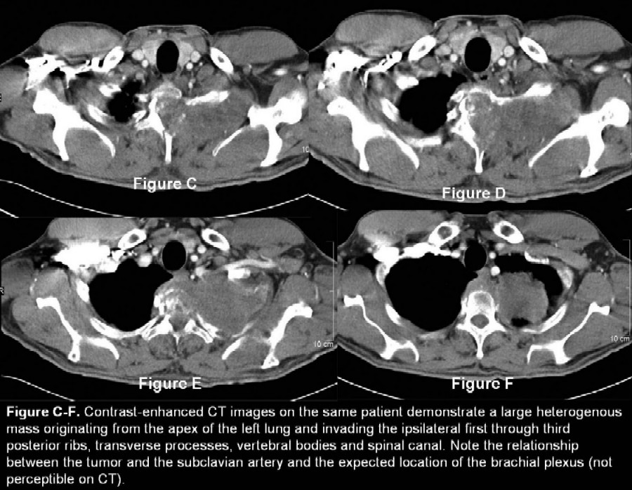
· Apical mass; bulk of which is extrathoracic (Fig. C-F)
· Chest wall invasion with associated posterior rib and or thoracic spine involvement (Fig. C-F)
· +/- Vascular invasion
o Subclavian vein
o Subclavian artery
o Superior vena cava
· +/- Tracheal invasion
· +/- Scalene muscle invasion
· +/- Lymphadenopathy
· +/- Metastatic pulmonary nodules
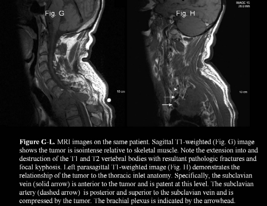
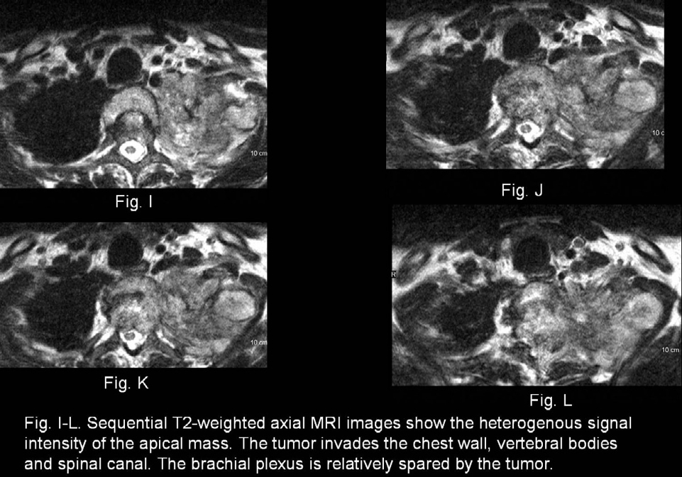
MRI
· Imaging method of choice
· Better delineates the relationship between the tumor and the adjacent neurovascular structures including the brachial plexus
o Subclavian artery involved more frequently than the subclavian vein due to its more posterior location and close association with the lung parenchyma.
· Chest wall invasion and extension into the vertebral bodies, spinal canal, and adjacent musculature
Staging
By virtue of their location and chest wall involvement, Pancoast tumors are typically defined as at least T3 lesions. Invasion of the brachial plexus, mediastinum, or spine, elevates their classification to T4. Thus, by definition Pancoast tumors are at minimum stage III tumors at the time of presentation.
Treatment
o Workup:
· Typical NSCLC workup:
· CT chest and abdomen
· CBC, LFTs, electrolytes
· Bone scan
· [FDG]-PET imaging or mediastinoscopy to evaluate for mediastinal node involvement for triaging patients to operative versus non-operative treatment
· US or CT guided percutaneous biopsy
· MRI of the chest and MRA to evaluate tumor extent and surgical resectability.
Treatment:
· Surgery:
· Lobectomy rather than wedge resection has been shown to improve survival.
· Vascular and vertebral body involvement was once considered a contraindication to surgical intervention. However, as surgical technique and technology have evolved, these tumors may now undergo resection.
· Extensive involvement of the brachial plexus remains a contraindication to resection; however, Horner’s syndrome is not a contraindication.
· Metastasis to contralateral mediastinal lymph nodes is considered a contraindication for surgical resection.
· Radiation therapy
· External beam or brachytherapy generally used as multimodality therapy prior to surgery, which improves resectability, decreases tumor seeding at surgery, blocks lymphatic channels, and increases local control.
· Chemotherapy
· Historically utilized as palliative treatment in metastatic disease.
· Current treatment now involves chemotherapy as induction therapy prior to definitive surgery or radiation therapy or with concurrent radiation therapy.
· Concurrent chemotherapy and radiation therapy followed by surgery
· In one multi-center study and several single center studies, combined chemo and radiation prior to surgery has been shown to:
· Increase the rate of complete resection (92% vs. average 66% with conventional therapy of radiation prior to surgery).
· Decrease local recurrence rate (23% vs. 58%).
· Increase 2 yr survival (55% vs. 42%).
· Over 1/3rd of patients have complete pathologic responses, while an additional 30% have minimal microscopic disease upon resection.
· Thus, preoperative chemoradiotherapy has become the standard of care for patients with Pancoast tumors.
Prognosis
Pancoast tumor treated with chemoradiation therapy prior to surgery:Most recent data have shown:
· 2-year survival rates of 55% for all patients; 70% for those who underwent complete resection
· Median survival of 33 months and 5-year overall survival rate of 41% for all patients.In a third of surgical resections a complete resection is not achieved; these patients have survival rates similar to no resection at all.
· Recurrences
o Historically, local recurrences have been the most frequent type. With preoperative chemoradiation therapy, however, distant metastases now represent the majority of recurrences.
Caveats
· Pancoast tumor arises from the apex of the lung, the bulk of the tumor is extrathoracic, and the tumor principally invades the apical chest wall and or its associated neurovascular structures rather than the underlying lung parenchyma.
· By virtue of their location and chest wall involvement, Pancoast tumors are typically defined as at least T3 lesions.
· MRI is the staging imaging modality of choice. It better delineates the relationship between the tumor and the adjacent neurovascular structures including the brachial plexus.
· Extensive involvement of the brachial plexus remains a contraindication to resection; however, Horner’s syndrome is not a contraindication.
Selected Readings
1. Arcasoy, Selim M. " Superior pulmonary sulcus tumors and Pancoast's syndrome." The New England Journal of Medicine 337.19 (1997): 1370-376.
2. Archie, Victor C. " Superior sulcus tumors: A mini-review." The Oncologist (2004): 550-55.
3. Detterbeck, Frank C. "Changes in the treatment of Pancoast tumors." Annals of Thoracic Surgery 75 (2003): 1990-1997.
4. Siegel, B. A., and A. V. Proto, eds. Chest disease (fifth series) test and syllabus. Reston , Va : American College of Radiology, 1996. Case 15: Superior Sulcus Carcinoma (Pancoast Tumor)
Original case written by its authors at Virginia Commonwealth University and published at this address as part of a weekly teaching collection. Reproduced here as an archive.