Differential Diagnosis
- Pneumococcal pneumonia
- Mixed anaerobic infection
- Haemophilus influenzae pneumonia
- Staphylococcal pneumonia
Discussion
Background
Klebsiellae are ubiquitous in nature. In humans, they may colonize the skin, pharynx, gastrointestinal tract, sterile wounds and urine and may be regarded as normal flora in many parts of the gastrointestinal and biliary tract. Oropharyngeal carriage is associated with endotracheal intubation, impaired host defenses, and antimicrobial use. The primary pathogenic reservoirs are the gastrointestinal tract of patients and the hands of hospital personnel, the latter often responsible for nosocomial outbreaks. Klebsiella pneumonia usually results from aspiration of colonizing oropharyngeal microbes into the lower respiratory tract. Risk factors for Klebsiella pneumonia include alcoholism, diabetes mellitus, and chronic obstructive lung disease.
Etiology
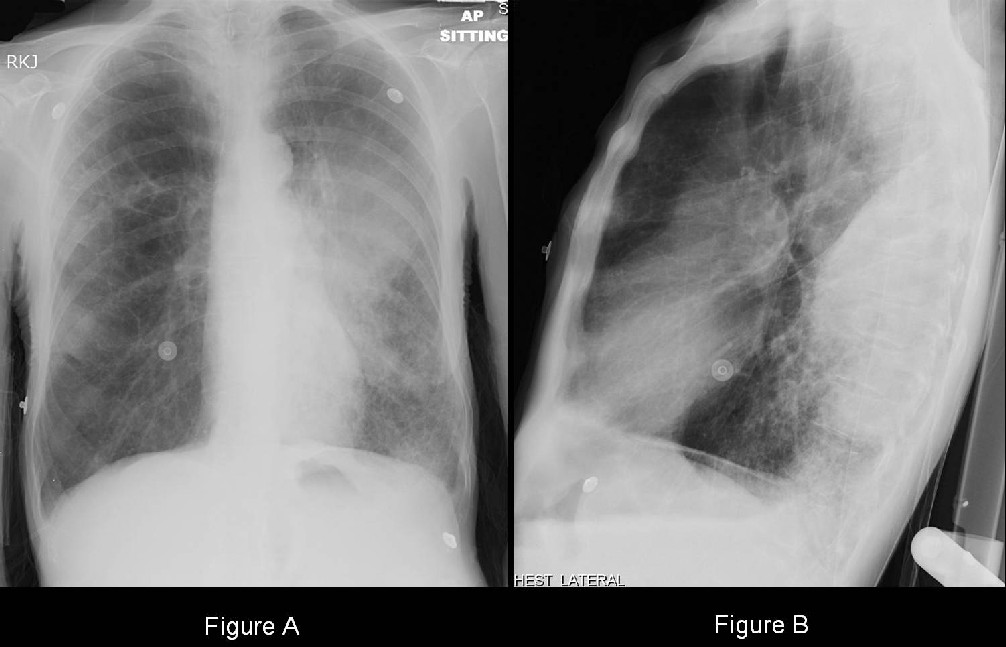
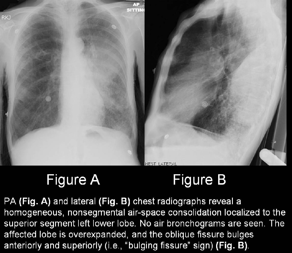
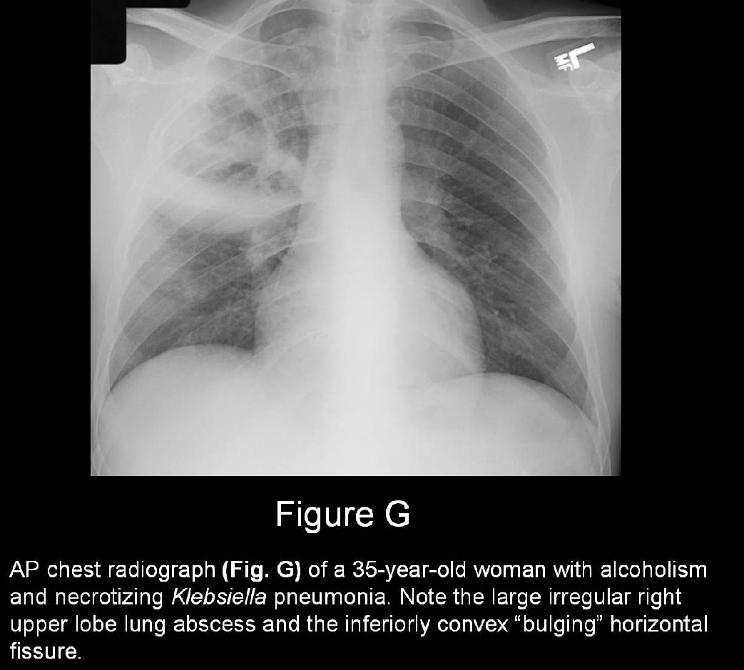
Klebsiella pneumonia is caused by the gram-negative bacterium Klebsiella pneumoniae. K. pneumoniae causes approximately 5% of community-acquired pneumonia and up to 30% of nosocomial pneumonia. The extensive clinical use of broad-spectrum antibiotics in hospitalized patients has led to an increased carriage of Klebsiella as well as the evolution of multi-drug-resistant strains (MDRS). Lung infection by K. pneumoniae is often associated with the production of a large volume of inflammatory exudate. The exudate may infiltrate the entire affected lobe, which enlarges, over-expands, and causes the abutting fissure to bulge toward the unaffected lobe (“bulging fissure” sign). Lung infection may also be associated with necrosis, tissue destruction and hemorrhage sometimes producing thick, bloody, mucoid sputum described as currant jelly.
Clinical Findings
Klebsiella pneumonia primarily affects persons with alcoholism (66%) and the debilitated (ICU and nursing home patients). Additional risk factors include indwelling central venous catheters and feeding tubes. Affected patients present with the sudden onset of fever, rigors, dyspnea, pleuritic chest pain, and a productive cough with the consistency of currant jelly Prostration and hypotension may occur. The white blood count may be elevated, diminished, or normal. Klebsiella pneumonia may be complicated by multicentric abscesses, cavitation, pulmonary gangrene, bronchiectasis, and empyema. Chronic pneumonia may be associated with interstitial fibrosis, organizing pneumonia, bronchiolitis, and necrotizing bronchitis.
Imaging Findings
Radiography
- Usually involves one of the upper lobes; however involvement of lower lobes is not uncommon (Fig. G).
- Homogeneous, nonsegmental, lobar consolidation (Figs. A and B).
- Lobar expansion; “bulging fissure” sign (Figs. B and Fig. G).
- Lung abscess (es); occur in up to 50% cases (Fig. G).
- Pulmonary gangrene
- Begins as a lobar consolidation; usually in the upper lobes.
- Coalescence of intrinsic lucencies to form a large cavity.
- “Mass within a mass” or “air crescent” signs secondary to sloughed lung parenchyma or lung necrosis.
- Pleural effusion (70%) and/or empyema.
CT
- Necrotizing pneumonia
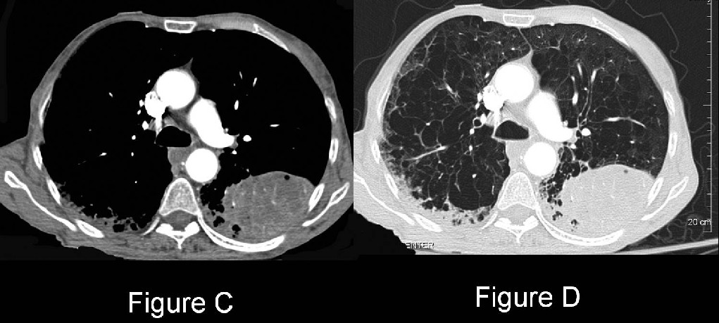
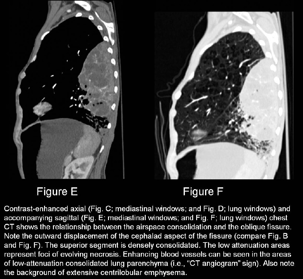
- Enhancing consolidations and poorly marginated low-attenuation areas with or without small air-containing cavities (Figs. C-F).
- Scattered enhancing linear branching structures representing pulmonary vessels in atelectatic or consolidated lung (e.g., “CT angiogram” sign) (Figs. C-F).
- Centripetal resolution from the periphery to the center with residual fibrosis.
- Pulmonary gangrene
- Coalescence of multiple small abscesses into a large cavity containing sloughed or necrotic lung.
- Narrowed or obliterated feeding bronchus impeding drainage of necrotic and infected lung.
- Large-vessel thrombosis.
- Pleural effusion and/or empyema.
Management
Community-acquired pneumonia
- Third-generation cephalosporins or quinolones and/or combination therapy with aminoglycosides.
- Antibiotic therapy should be continued for at least 14 days.
- Clinical and radiologic surveillance for surgically treatable complications
- Pulmonary gangrene.
- Lung abscess (es).
- Empyema.
Nosocomial pneumonia
- Imipenem, third-generation cephalosporins, quinolones, or aminoglycosides used alone or in combination.
- Always confirm organism susceptibility.
- Antibiotics should be continued for at least 14 days.
Prognosis
- Klebsiella pneumonia is a severe illness with a rapid onset; mortality approaches 50% even with antimicrobial therapy.
- Mortality may approach 100% for those persons with predisposing alcoholism and bacteremia.
PEARLS
- Klebsiella is among the top 8 nosocomial pathogens in hospitals. Any organ system may be affected, but respiratory and urinary tract infections predominate.
- Because of the high prevalence of pneumococcal pneumonia in most communities, the majority of patients with pneumonia manifesting with a “bulging fissure” sign are infected with S. pneumoniae rather than with K. pneumoniae.
- Cavitation, especially in the presence of a unilateral necrotizing pneumonia, strongly supports Klebsiella as the possible etiology.
Suggested Readings
- Parker MS, Rosado-de-Christenson ML, Abbott GF. Pulmonary Infections and Aspiration Pneumonia: Common Bacterial Pneumonias. In: Teaching Atlas of Chest Imaging. New York, New York: Thieme; 2006: 223-226.
- Schmidt AJ, Stark P. Radiographic findings in Klebsiella (Friedländer’s pneumonia): the bulging fissure sign. Semin Respir Infect 1998; 13:80-82.
- Travis WD, Colby TV, Koss MN, Rosado-de-Christenson ML, Müller NL, King TE Jr. Lung infections. In: King DW, ed. Atlas of Nontumor Pathology: Non-Neoplastic Disorders of the Lower Respiratory Tract. First series, fascicle 2. Washington, DC: American Registry of Pathology; 2002:549-550.
Original case written by its authors at Virginia Commonwealth University and published at this address as part of a weekly teaching collection. Reproduced here as an archive.