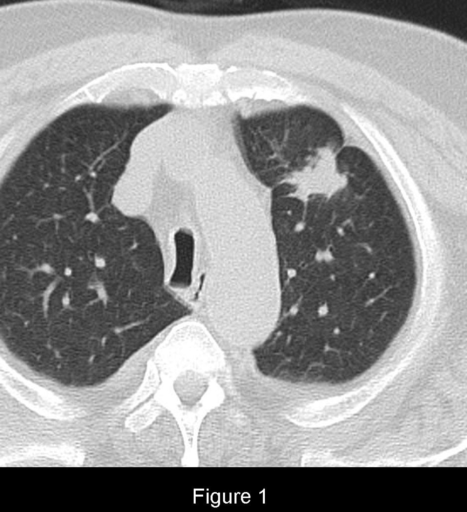
Radiologic Findings Single unenhanced chest CT image (Fig. 1) (lung windows) through the upper lung zones shows a 4.0 cm polylobulated mass in the peripheral anterior segment of the left upper lobe. The mass is surrounded by aerated lung but does demonstrate irregular borders and a pleural tail. Diagnosis: T2a N0 M0 Stage IB Adenocarcinoma of the Lung
Differential Diagnosis
Not Applicable
Discussion
In 2009, based on proposals from the International Association for the Study of Lung Cancer and the Lung Cancer Staging Project, the International Union Against Cancer (IASLC) and the American Joint Committee on Cancer (AJCC) released the revised 7th edition of the TNM classification for Non-Small Cell Lung Cancer. The revisions were based on a large database derived from 45 institutions and 20 nations under the auspices of the IASLC. Most of the revisions were directed towards the T (tumor) and M (metastases) descriptors and did not affect the N (nodal) descriptors.
T (tumor)-reflects the greatest dimension or size of the lesion as well as its location in the thorax relative to the airways and its degree of invasiveness with respect the pleura, chest wall, and mediastinum. Under the revised system, the T of the lesion is question is designated based on the following size criterion:
T1a T1b T2a T2b T3 ≤2.0 cm > 2.0 ≤ 3.0 cm >3.0 ≤ 5.0 cm >5 cm ≤ 7 cm >7 cm The mere presence of a pleural tail does not indicate visceral pleural invasion. As a radiologist staging a patient, we are always obligated to down stage a patient not upstage the patient.
There are additional “T” criterion applied with respect to a given tumor’s location relative to the airway and its degree of invasiveness with respect to the chest wall, pleura, and mediastinal structures. These latter criteria are beyond the scope of today’s discussion. The reader is referred to the Selected Readings at the end of today’s case discussion to learn more.
As the lesion in our test case is >3 cm but ≤ 5 cm and surrounded by aerated lung, its clinical designation is T2a.
In the absence of nodal or metastatic disease, (i.e., N0; M0) under the revised system, this lesion’s clinical stage would be Stage IB
N0 N1 N2 N3 T1a IA IIA IIIA IIIB T1b IA IIA IIIA IIIB
T2a IB IIA IIIA IIIB T2b IIA IIB IIIA IIIB T3 IIB IIIA IIIA IIIB T4 IIIA IIIA IIIB IIIB M1a IV IV IV IV M1b IV IV IV IV
Selected Readings
- Diederich S. Lung Cancer Staging Update: The Revised TNM Classification. Cancer Imaging 2010; 10: Spec no A: S134-135.
- Klingerman S, Abbott G. A Radiologic Review of the New Classification for Lung Cancer. AJR 2010; 194(3): 562-573.
- Rami-Porta R, Crowley JJ, Goldstraw P. The Revised TNM Staging System for Lung Cancer. Ann Thorac Cardiovasc Surg 2009; 15(1): 4-9.
- www.iaslc.org
can you purchase viagra online where to purchase viagra trusted viagra sites generic ed medication buy amoxicillin cheap generic viagra online amoxicillin buy can you purchase viagra online where to purchase viagra trusted viagra sites generic ed medication buy amoxicillin cheap generic viagra online amoxicillin buy can you purchase viagra online where to purchase viagra trusted viagra sites generic ed medication buy amoxicillin cheap generic viagra online amoxicillin buy can you purchase viagra online where to purchase viagra trusted viagra sites generic ed medication buy amoxicillin cheap generic viagra online amoxicillin buy
Original case written by its authors at Virginia Commonwealth University and published at this address as part of a weekly teaching collection. Reproduced here as an archive.