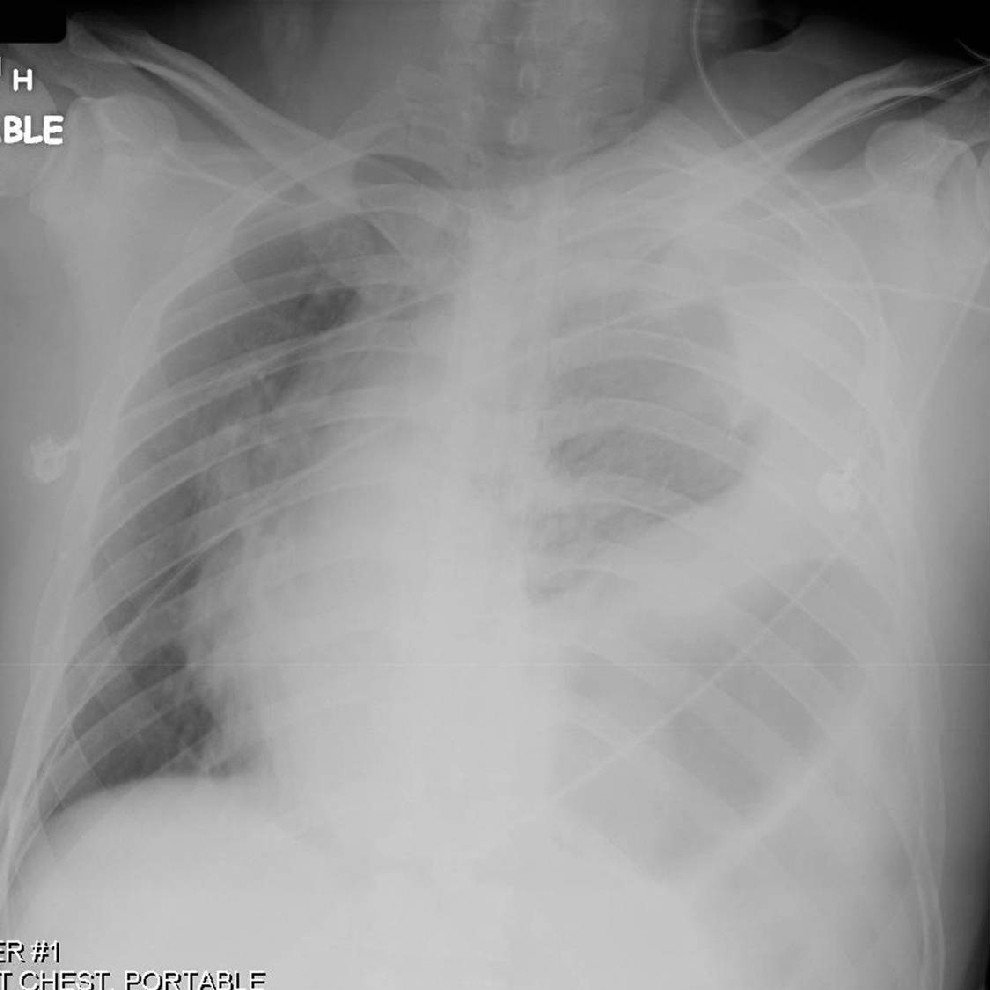
AP chest radiograph shows marked rightward displacement of the cardiomediastinal silhouette by herniated hollow viscera into the left thorax. A left-sided hemothorax extends into the apex. Multiple left-sided rib fractures are present. Diagnosis: Acute traumatic rupture left diaphragm
Differential Diagnosis
- Eventration or elevation of the diaphragm
- Diaphragmatic paralysis
Discussion
Background
Most diaphragmatic injuries are caused by penetrating trauma. Approximately 15% of stab wounds and 45% of gunshot wounds to the lower chest are complicated by diaphragmatic injuries. Diaphragmatic injuries should be suspected in any penetrating trauma victim with wounds below the 4th anterior, the 6th lateral, and the 8th posterior intercostal spaces (“4-6-8 rule”). The incidence of diaphragmatic injury among blunt trauma victims varies from 0.8 to 8.0% and is more frequently observed with abdominal as opposed to thoracic trauma. Left-sided injuries are approximately 3 times more common than right-sided injuries, however, as many as 4.5% of blunt trauma victims sustain bilateral diaphragmatic injuries. Most diaphragmatic tears are more than 10 cm in length and involve the muscular posterior or posterolateral diaphragm.
Etiology
A sudden increase in either intra-abdominal or intra-thoracic pressure against a fixed diaphragm accounts for most blunt diaphragmatic injuries. Other postulated mechanisms of injury include shearing stress on a stretched diaphragm and avulsion of the diaphragm from its points of attachment.
Clinical Findings
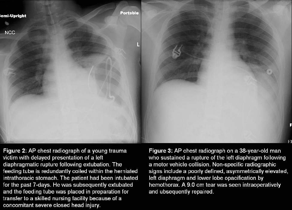
Acute diaphragmatic injury should be considered in patients who sustain significant abdominal or thoracoabdominal trauma and present with dyspnea or respiratory distress. The majority of affected trauma patients (<94%) have concomitant injuries. Associated injuries often include hepatic (16%) and/or splenic lacerations (48%); rib fractures (52%); pelvic fractures (52%); and closed head injuries (32%). Left-sided diaphragmatic injuries are associated with herniation of subdiaphragmatic viscera into the chest. The stomach and colon are the most commonly herniated organs. As many as three quarters of affected patients have herniation of other intraabdominal organs (e.g., omentum, spleen, kidney, and pancreas). Diaphragmatic injuries may go unrecognized in patients on positive pressure ventilation only declaring their presence following extubation (Fig. 2). Delayed herniation may be complicated by bowel strangulation. Intrathoracic splenosis may manifest as sequelae of remote diaphragmatic and splenic injuries on imaging studies decades later, and should not be confused with neoplastic disease.
Imaging Findings
Radiography
Initial chest radiography is diagnostic in 27-60% of left-sided diaphragmatic injuries, but in only 17% of right-sided injuries.
- May initially appear normal.
- Non-specific findings may include:
- Elevated asymmetric, poorly visualized, or irregular-appearing diaphragm (Figs. 1-3).
- Hemothorax, pneumothorax, and hemopneumothorax (Fig. 1 and Fig. 3).
- Lower lobe opacification in the presence of an elevated diaphragm (Fig. 3).
- Contralateral mediastinal shift in the absence of a pneumothorax or large effusion (Fig. 1).
- Persistent contralateral mediastinal shift despite the presence of a thoracostomy tube.
- Lower rib fractures.
- More specific findings may include:
- An abnormal or U-shaped course of the nasogastric or enteric tube (Fig. 2).
- Herniation of abdominal contents into the thorax (Figs. 1 and Fig. 2).
Ultrasonography
Scanning is performed in the oblique transverse subxiphoid plane at the midline to obtain comparative images of both hemidiaphragms. Transducer is then positioned in each subcostal area, and each hemidiaphragm is scanned separately in the coronal plane.
- Permits direct visualization of the diaphragm.
- More useful in the evaluation of right-sided injuries.
- Stomach/bowel gas limits evaluation of left-sided injuries.
- Normal diaphragm demonstrates continuous echogenic lines; injured diaphragm shows focal disruptions or interruptions of diaphragmatic echoes at the injury site.
- Diminished or absence of the expected respiratory excursion of the ruptured diaphragm.
MDCT
Blunt Trauma:
S ensitivity- 50-100%; specificity-86-100%
Imaging Findings
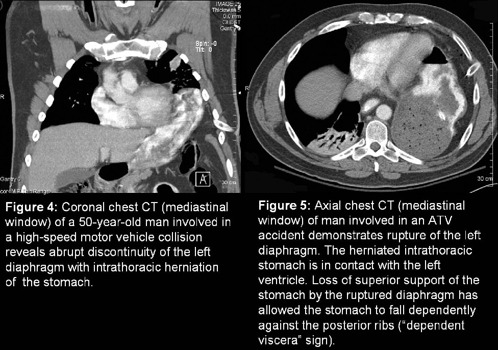
- Abrupt discontinuity of the diaphragm (73-82%) with or without herniation of the stomach or other viscera into the thorax (Fig. 4).
- “Absent diaphragm” sign
- Non-visualization of the diaphragm in an area where it does not contact another organ and should otherwise is seen.
- “Dependent viscera” sign
- Loss of the superior support of the liver, stomach, and or bowel allowing them to fall dependently against the posterior ribs (Fig. 5).
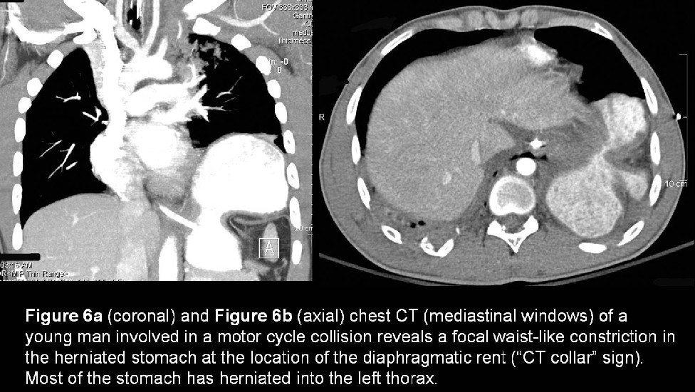
- “CT collar” sign
- Waist-like or focal constriction of herniated viscera, stomach or bowel the diaphragmatic defect (Fig. 6).
- Visualization of peritoneal fat, bowel, or viscera lateral to the lung or diaphragm or posterior to the diaphragmatic crus.
- “Hump” sign
- Variant of the “collar sign”
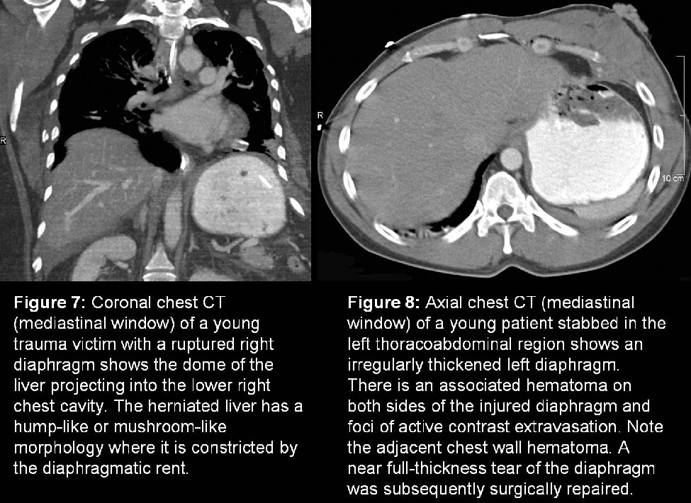
- Rounded portion of herniated liver through the diaphragm forms a hump-shaped mass (Fig. 7).
- May also manifest as a mushroom-like mass in the right hemithorax where the herniated liver is constricted by the tear.
- “Band” sign
- Linear lucency across the liver along the torn edges of the hemidiaphragm.
- Irregularly thickened diaphragm
- Highly suggestive of diaphragmatic rupture in the absence of retroperitoneal contusion, but does not distinguish between injury requiring surgical repair and partial-thickness tears.
- Concomitant proximity injuries of the ribs, liver and/or spleen.
Penetrating Trauma:
Sensitivity-88%; specificity-82%
- Most accurate sign is the presence of a contiguous injury on either side of the diaphragm in single-entry penetrating trauma (Fig. 8).
MRI
- Primary role is in nonacute or difficult cases (e.g., eventration/elevation versus injury).
- Both cardiac and respiratory gating should be used to minimize motion artifacts.
- TIWI sagittal and coronal images delineate the left diaphragm as a low signal intensity curvilinear band of soft tissue outlined by higher signal intensity abdominal and mediastinal fat.
- Evaluation of the right diaphragm is more problematic.
Treatment
- Small diaphragmatic lacerations
- Laparoscopy is an alternative to open repair.
- Open laparotomy for large (> 10 cm) tears adjacent to or near the esophageal hiatus.
- Most injuries can be repaired primarily.
- Synthetic mesh (e.g., polypropylene, Dacron) occasionally required for large tears.
- Centrally located injuries
- Posterolateral injury of the right diaphragm
- Best approached through the chest as the liver obscures an abdominal approach.
- Lateral injuries near the chest wall
- May require reattachment of the diaphragm to the chest wall by encirclement of the ribs with suture material.
Prognosis
- Mortality approaches 30%.
- May be adversely affected by
- Associated abdominal and thoracic injuries.
- Delays in diagnosis and surgical repair; bowel strangulation.
Caveats
- Positive-pressure ventilation can prevent the herniation of bowel into the thorax, delaying the diagnosis of diaphragmatic injury in some cases until after the patient has been extubated.
- Associated atelectasis and or hemothorax may obscure visualization of diaphragmatic tears.
- Normal focal areas of discontinuity in the posterior diaphragm are seen in 6-11% of nontrauma patients (e.g., congenital Bochdalek defects) and in up to 35% of elderly persons. The presence or absence of concomitant proximity trauma is helpful in appropriately directing the diagnosis.
Suggested Readings
- Bagheri R, Tavasoli A, Sadrizadeh A, Rajabi Mashhadi M, Shahri F, Shojaeian R. The role of thoracoscopy for the diagnosis of hidden diaphragmatic injuries in penetrating thoracoabdominal trauma. Interact Cardiovasc Thorac Surg 2009; May 25. [Epub ahead of print].
- Bodanapally UK, Shanmuganathan K, Mirvis SE, Sliker CW, Fleiter TR, Sarada K, Miller LA, Stein DM, Alexander M. MDCT diagnosis of penetrating diaphragm injury. Eur Radiol 2009; Mar 31. [Epub ahead of print].
- Costantino M, Gosselin MV, Primack SL. The ABC’s of Thoracic Trauma Imaging. Semin Roentgenol 2006 Jul; 41(3):209-25.
- Gavelli G, Canini R, Bertaccini P, et al. Traumatic injuries: imaging of thoracic injuries. Eur Radiol 2002; 12: 1273-1294.
- Lomoschitz FM, Eisenhuber E, Linnau KF, Peloschek P, Schoder M, Bankier AA. Imaging of chest trauma: radiological patterns of injury and diagnostic algorithms. Eur J Radiol 2003; 48: 61-70.
- Primack SL, Collins J. Blunt nonaortic chest trauma: radiographic and CT findings. Emerg Radiol 2002; 9: 5-12.
Original case written by its authors at Virginia Commonwealth University and published at this address as part of a weekly teaching collection. Reproduced here as an archive.