Archived case 77 · Nov 19-Nov 26, 2010
Calcified Tuberculous Cervical Lymphadenitis (Scrofula)
Clinical Presentation Withheld
The question posed to readers
Describe the radiologic findings and the underlying etiology
Archived case 77 · Nov 19-Nov 26, 2010
Clinical Presentation Withheld
Describe the radiologic findings and the underlying etiology
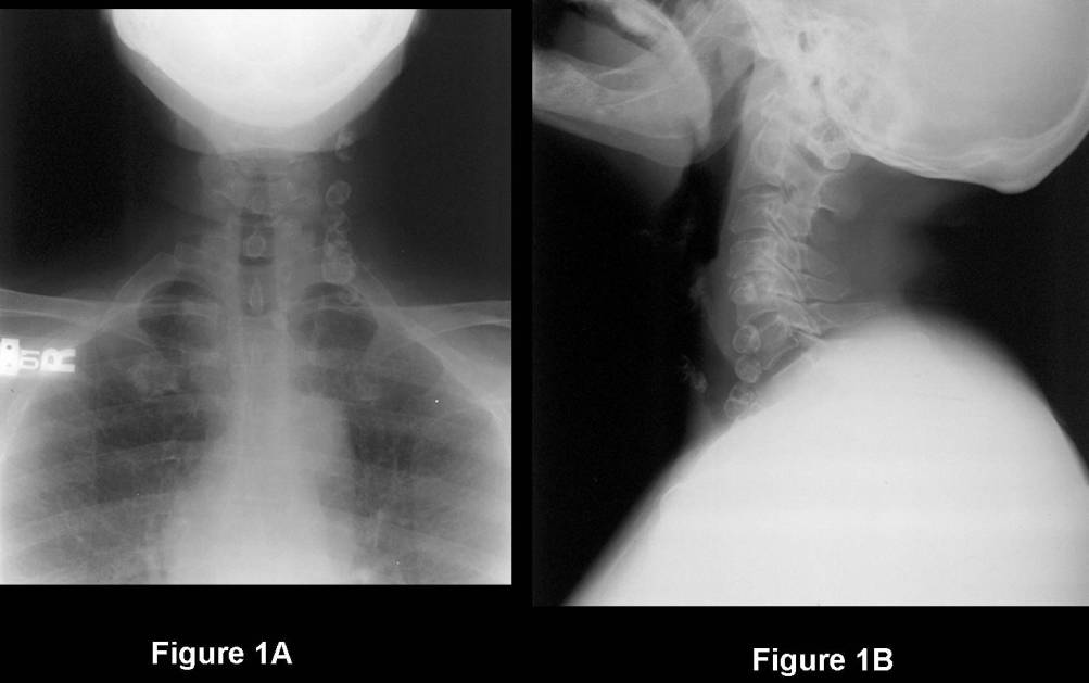
Calcified Tuberculous Cervical Lymphadenitis (Scrofula)
Differential Diagnosis
Granulomatous Infection or Inflammatory Processes of other Etiologies
Discussion
Tuberculous lymphadenitis or scrofula (after the Latin word “scrofa” for sow’s milk) was first described in Medieval times. Centuries latter discovered that the infection was caused by the ingestion of infected cow’s milk and most commonly from infection with Mycobacterium bovis.
Affected patients presented with swollen “glands” in the neck. For centuries, people believed that Kings had been given divine powers from God to rid people of this “curse” and that the simple touch by their royal hand would cure those affected. Large pompous ceremonies were often carried out during their “cleansing process”. In addition to the affected person and the King, a choir, ladies and gentlemen of the court, and a troop or two of Beefeaters participated in the ceremony. In fact, Charles II, King of England, Scotland, Ireland, one of the cleverest but most controversial of English Kings, is credited with having “Touched” more than 92,000 affected persons during his reign.
Etiology
Cervical lymphadenitis is the most common form of extra-pulmonary Tuberculosis.
Tuberculous infection of the neck may be caused by:
Present day tuberculous cervical lymphadenitis is more commonly caused by inhalation rather than ingestion. The bacterium spreads throughout the body and affects the cervical and supraclavicular lymph node chain.
Clinical Findings
Affected patients often present with “Rubbery” enlargement of cervical lymph nodes. There may be concomitant involvement of the lymph nodes in the supraclavicular fossa, axillae, and groin. The nodes may then ulcerate producing chronic draining sinuses and abscesses. Cervical lymphadenitis was relatively rare pre-AIDS epidemic. Since the HIV-AIDS epidemic, there has been a resurgence of both localized and systemic forms of tuberculosis. Mycobacterium scrofulaceum accounts for approximately 2% of mycobacterial infections in AIDS patients but also affects minorities, women, and children.
Immunocompetent Adults:
Immunocompetent Children:
Immunocompromised (HIV +) Children:
Differential Diagnosis Cervical Lymphadenitis-Immunocompetent Patients
Differential Diagnosis Cervical Lymphadenitis-Immunocompromised (HIV +)
Treatment
Immunocompetent
Immunocompromised
Selected Readings
Filed under: Radiology, Medicine/Pulmonary
Original case written by its authors at Virginia Commonwealth University and published at this address as part of a weekly teaching collection. Reproduced here as an archive.