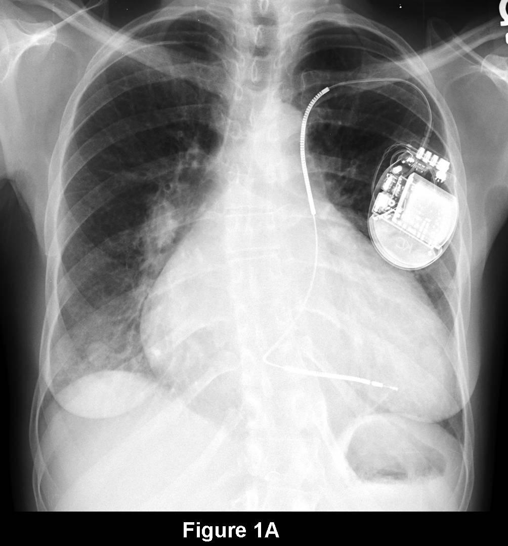
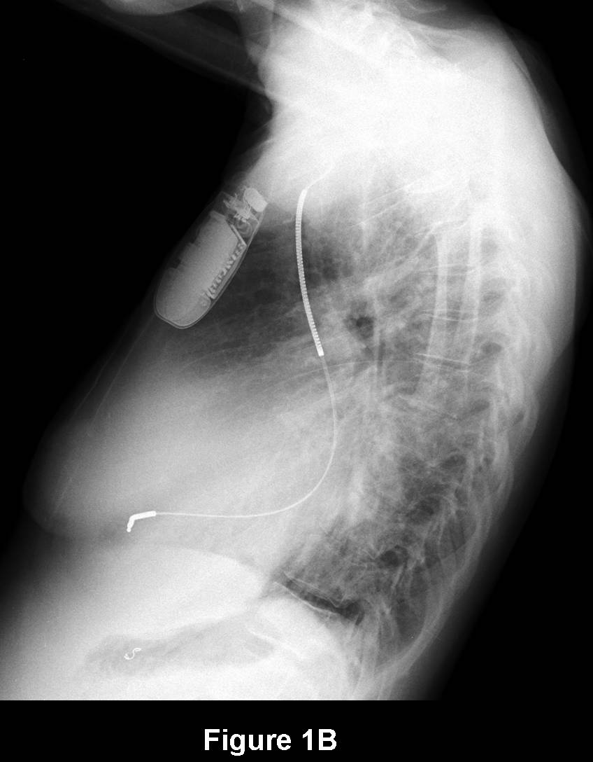
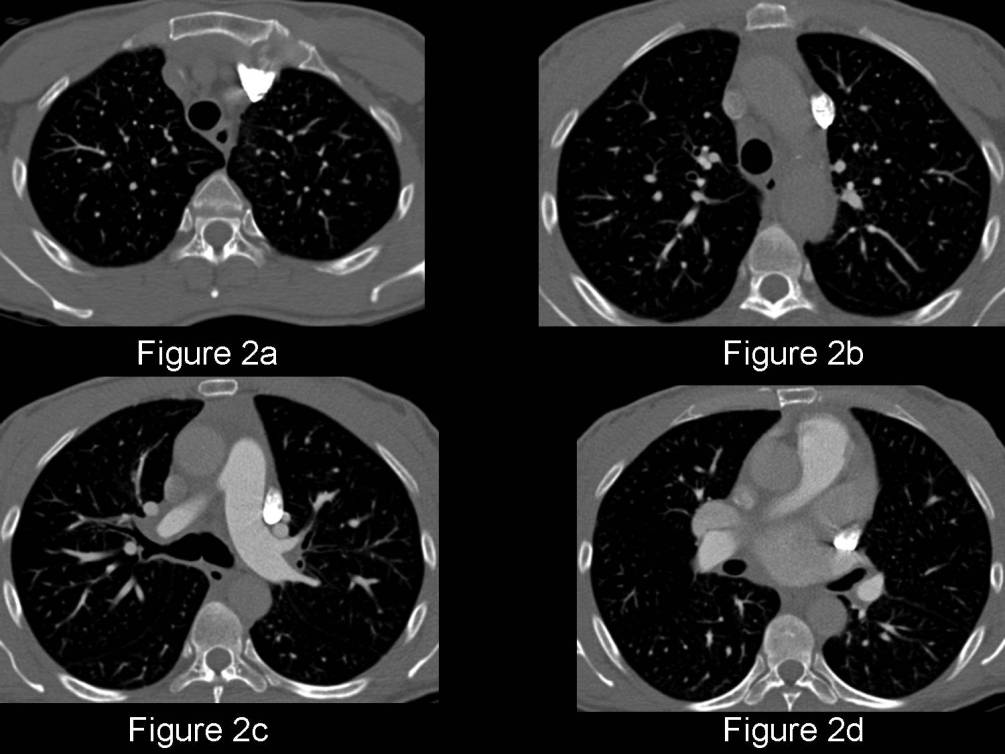
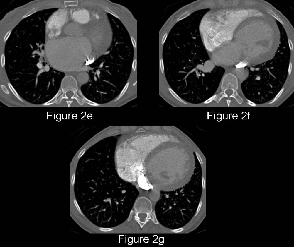
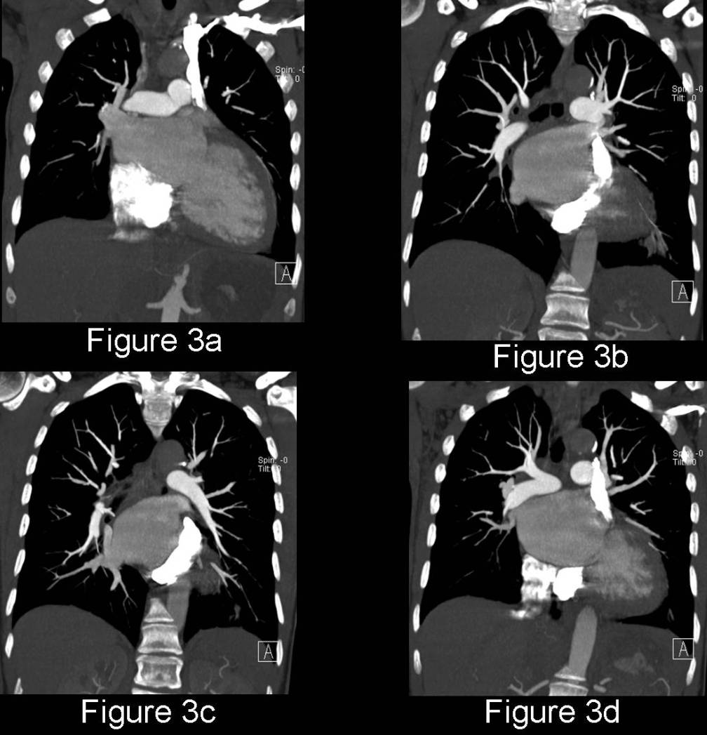
Radiologic Findings PA (Fig. 1A) and lateral (Fig. 1B) chest radiographs demonstrate an enlarged cardiomediastinal silhouette compatible with the patient’s known underlying cardiomyopathy. There is currently no evidence of cardiac decompensation or volume overload. A left subclavian transvenous approach right ventricular implantable cardioverter-defibrillator (ICD) is present. It follows an unusual course along the left heart border, but not an unexpected course in the setting of a persistent left superior vena cava, coursing into the coronary sinus, right atrium and then on into the right ventricular chamber. Contrast-enhanced axial (Fig. 2A-G) and coronal (Fig. 3A-D) (modified mediastinal window) confirm the ICD follows the course of a persistent left superior vena cava. The anomalous vena cava courses vertically along the left superior mediastinum and continues caudally coursing posterior to the left atrial appendage and left atrium and anterior to the left superior pulmonary vein and then enters the coronary sinus. Note the right superior vena cava (Images courtesy of Daniel A Henry, MD, VCU Medical Center). Diagnosis: Persistent Left Superior Vena Cava (PLSVC)
Differential Diagnosis
None
Discussion
A relatively common anomaly, persistence of the LSVC occurs in approximately 0.3% of the general population. The most frequent form of anomalous venous return to the heart, PLSVC is typically diagnosed in asymptomatic adults undergoing chest radiography for unrelated reasons. However, given the fact that PLSVC is associated with abnormalities of impulse formation and conduction, and that different techniques are required to achieve optimal electrical functioning when placing pacemaker or defibrillator devices; recognition of a persistent LSVC is of clinical importance. Of further importance, PLSVC also can complicate central venous line placement and cardiac/thoracic surgery procedures.
Prevalence
- Occurs in 0.3% of general population
- Prevalence in patients with congenital cardiac anomalies ranges from 2.8-4.3%
- Given the association with disturbances of cardiac impulse formation/conduction, one would expect a higher prevalence over the general population, though Biffi, et al reported finding prevalence approximately equal to that of the general population in their experience.
Etiology/Anatomy
- Early in fetal life, the sinus venosus has three parts (Right horn, transverse, and left horn) that drain three veins (omphalomesenteric, umbilical, and common cardinal veins)
- The anterior cardinal veins drain the upper body and the right and left anterior cardinal veins ultimately join to form left brachiocephalic vein
- Normally the left anterior cardinal vein involutes (ligament of Marshall). Failure to involute results in PLSVC
- Rarely, even in cases of PLSVC, the right anterior cardinal vein involutes. This results in absent right SVC seen in 10-33% of patient with PLSVC
- PLSVC arises at the junction of the left jugular and subclavian veins
- Usually, the PLSVC drains into the right heart via the coronary sinus
PEARLS
- Separate from associated congenital heart disease, the prognosis for PLSVC is excellent
- Once PLSVC is identified, interventional cardiology may place wire through PLSVC as in this case, through the usual right SVC approach, or may convert to epicardial implantation of leads
- PLSVC necessitates special surgical techniques to avoid flooding cardiac chambers during open heart surgery. Ligation of PLSVC in patients lacking right SVC or Left brachiocephalic vein connection to SVC leads to venous engorgement.
- Though normally draining into the right atrium, drainage into the left atrium can occur via fenestrations between coronary sinus and the left atrium or by atrial septal defect (ASD) at the mouth of coronary sinus.
Selected Readings:
- Biffi, M. et al. Left Superior Vena Cava Persistence in Patients Undergoing Pacemaker or Cardio-Defibrillator Implantation: A 10-Year Experience. Chest. 2001; 120. 139-144.
- Parker MS, Rosado-de-Christenson ML, Abbott GF. Developmental Anomalies: Anomalies of the Aorta and Great Vessels. In: Teaching Atlas of Chest Imaging. 2006. 66-70. Thieme, New York.
Original case written by its authors at Virginia Commonwealth University and published at this address as part of a weekly teaching collection. Reproduced here as an archive.