Archived case 74 · Oct 29-Nov 5, 2010
Scimitar Syndrome
33-year-old woman with positive PPD
The question posed to readers
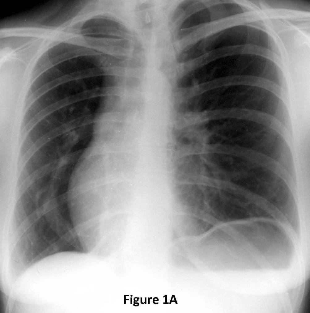
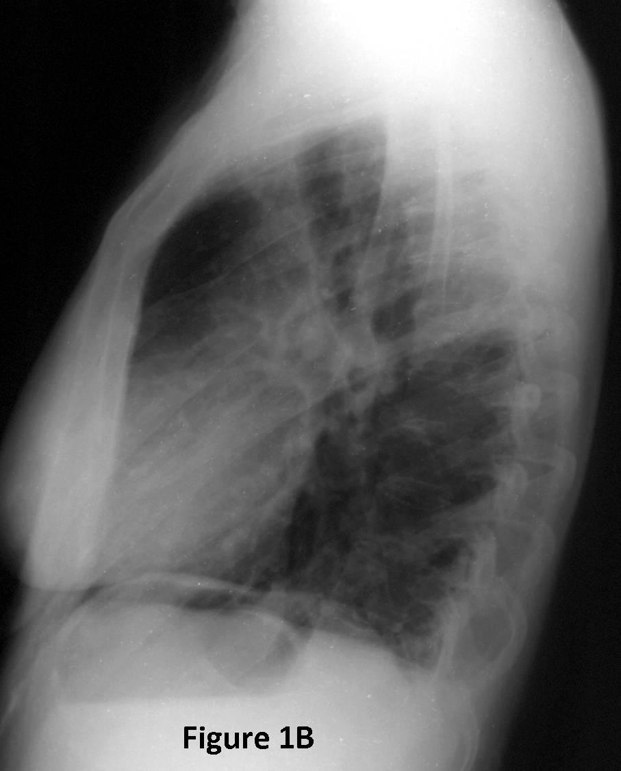
What is the pertinent radiologic finding? What is your diagnosis?
Archived case 74 · Oct 29-Nov 5, 2010
33-year-old woman with positive PPD
What is the pertinent radiologic finding? What is your diagnosis?
Scimitar Syndrome
Differential Diagnosis
Discussion
Scimitar syndrome is a rare congenital anomaly of the right lung and pulmonary vasculature of unknown etiology and is also known as pulmonary venolobar syndrome and hypogenetic lung syndrome. It characteristically involves the right lung and may be associated with a variety of cardiopulmonary anomalies. “Scimitar” is a descriptive term that refers to the curvilinear or arcuate course followed by the anomalous draining pulmonary vein, which has been likened to the shape of a Turkish sword called a scimitar. The anomalous vein(s) may drain all or only a portion of the right lung and typically drains into the inferior vena cava. In other cases the anomalous draining vein drains into hepatic circulation, azygos vein, coronary sinus or right atrium. Associated findings may include: hypoplasia of the right pulmonary artery, interruption of the IVC, duplication, eventration or partial absence of the right hemidiaphragm, and tracheal trifurcation. The hypogenetic right lung may also demonstrate anomalous segmentation, or bronchial branching.
Clinical Findings
Scimitar syndrome may be diagnosed in infancy because of the associated symptomatic congenital cardiovascular disease (25-50%). Associated congenital cardiac anomalies include: atrial septal defect, ventricular septal defect, patent ductus arteriosus, tetralogy of Fallot, coarctation of the aorta, hypoplasia of the left heart, and various endocardial cushion defects. Some patients may present with hemoptysis and/or recurrent pulmonary infection. Approximately 50% of patients are asymptomatic and diagnosed incidentally on chest radiography acquired for unrelated reasons. Women are slightly more often affected with a female-to-male ratio of 1.4:1.
Imaging Findings
Radiography
MDCT / MRI
Treatment
Prognosis
Selected Readings
Filed under: Radiology, Medicine/Pulmonary
Original case written by its authors at Virginia Commonwealth University and published at this address as part of a weekly teaching collection. Reproduced here as an archive.