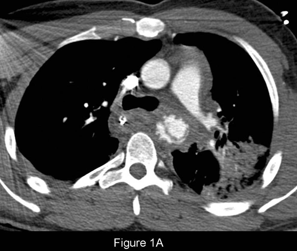
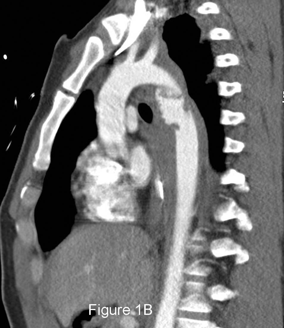
Contrast-enhanced axial CT (mediastinal window) (Fig. 1A) demonstrates gross irregularity in the descending thoracic aorta with an intimal flap extending from 2:00-11:00. Note the concomitant mediastinal hemorrhage and left upper lobe pulmonary contusion. The sagittal oblique MIP CT image (mediastinal window) (Fig. 1B) reveals the contour abnormality involves the entire circumference of the descending thoracic aorta beginning just distal to the ligamentum arteriosum and extends several centimeters in length. Again, note the extensive mediastinal hemorrhage. Diagnosis: Acute Post-Traumatic Aortic Injury (ATAI) with Pseudoaneurysm
Differential Diagnosis
None
Discussion
ATAI from blunt trauma is a substantial cause of morbidity and mortality, occurring in approximately 0.5%–2% of all non-lethal motor vehicle collisions (MVC) and 10%–20% of all high-speed deceleration fatalities. MDCT has emerged as the definitive screening modality for both diagnosis and exclusion of ATAI and injury of its great vessels without the need for conventional aortography or transesophageal echosonography. The latter are now typically only used in the infrequent setting of equivocal MDCT findings. In fact, the increased use of MDCT has led to the recognition and diagnosis of more subtle vascular injuries, which heretofore likely went undiagnosed, often with little or no surrounding peri-branch vessel or para-aortic hematoma. ATAI may occur anywhere along the thoracic aorta:
- Aortic Isthmus - within 2 cm of left subclavian artery origin; most common site (Fig. 1A and Fig. 1B)
- Aortic root and ascending aorta - 5%–14% of autopsies with aortic injuries; rarely seen at MDCT presumably because of its lethal nature
- Aortic arch and branch vessel injuries - less common. Isolated branch vessel injuries are more common than aortic arch injuries (<4% blunt chest trauma victims), but may be occur in combination with ATAI in 0%–45% of patients. Brachiocephalic and common carotid arteries are the most commonly involved vessels (66%–90%).
- Mid and distal descending thoracic aorta - 1%–12% of autopsies with aortic injuries.
- Minimal aortic injuries - only affect the intima and are estimated to occur in 10% of patients with ATAI; being encountered with increasing frequency. Nearly half of such cases show no abnormality at conventional angiography.
Clinical Findings
Clinical signs and symptoms are nonspecific and insensitive for the diagnosis and exclusion of ATAI. Most patients have no clinical signs of aortic injury until they suddenly become hemodynamically unstable. Symptoms induced by stretching of the mediastinal connective tissues by hemomediastinum include: interscapular pain, dyspnea, and hoarseness. Clinical signs of ATAI are absent in one-third of patients but when present include “pseudocoarctation syndrome”, external chest wall injuries, paraplegia, systolic murmur, and initial chest tube output > 750 mL of blood.
MDCT Imaging Features
MDCT features of aortic and great vessel injury are categorized as direct or indirect. The most specific direct signs are intimal flap and luminal thrombus or debris (100%) whereas irregular aortic contour or pseudoaneurysm is the most sensitive (100%).
Direct Signs (includes any of the following):
- Pseudoaneurysm
- Intraluminal flap
- Focal contour abnormality
- Abrupt aortic caliber change
- Coarctation
- Intraluminal thrombus or debris on the lacerated aortic wall
- Active contrast extravasation from injured aorta
Indirect Signs (includes any of the following):
- Subtle contour anomalies
- Hemomediastinum
- Peribranch vessel and/or peri-aortic blood
- Typically accompany direct signs but may occur in isolation
Treatment
Open Thoracotomy
- Left posterolateral thoracotomy
- Injured segment is resected; Dacron graft placed
- Post-operative paraplegia: 10%
- Mortality rate: 15-50%
Endovascular Stent-Graft Repair
- Obviates open repair, single lung ventilation, aortic cross-clamping, cardiopulmonary bypass, systemic anticoagulation
- Procedural planning with MDCT critical for technical success
- Morbidity: 0%–20%
- Limitation: Lack of small-caliber devices for use in young patients or those with small diameter aorta
Prognosis
- ATAI: high mortality rate; immediately fatal in 80%–90% of cases
- Substantial increase in mortality following ATAI in patients with concomitant injuries of the head, chest, and abdomen
- Prompt recognition and treatment is critical for long-term survival . If detected in a timely manner, 60%–80% of patients with ATAI reaching the hospital alive will survive following definitive therapy.
Selected Readings
- Creasy JD, Chiles C, Routh WD, Dyer RB. Overview of Traumatic Injury of the Thoracic Aorta. RadioGraphics 1997; Jan-Feb; 17(1):27-45.
- Fishman JE, Nuñez D Jr, Kane A, Rivas LA, Jacobs WE. Direct versus Indirect Signs of Traumatic Aortic Injury Revealed by Helical CT: Performance Characteristics and Interobserver Agreement. AJR Am J Roentgenol 1999; 172(4):1027-1031.
- Malhotra AK , Fabian TC, Croce MA, Weiman DS, Gavant ML, Pate JW. Minimal Aortic Injury: A Lesion associated with Advancing Diagnostic Techniques. J Trauma 2001 ; 51 : 1042 –1048.
- Steenburg SD , Ravenel JG, Ikonomidis JS, Sch o nholz C, Reeves S. Acute Traumatic Aortic Injury: Imaging Evaluation and Management. Radiology 2008; 248:748-762.
Original case written by its authors at Virginia Commonwealth University and published at this address as part of a weekly teaching collection. Reproduced here as an archive.