Thoracic Imaging Archive
34 language editions

Archived case 66 · Aug 30-Sep 6, 2010

Prominent Right 3rd Intercostal-Bronchial Artery Trunk; No Vascular Injury

28-year-old patient involved in a motor vehicle collision with complicating ejection and roll-over
Images

Radiograph 1 from archived case 66
Figure 1
Radiograph 2 from archived case 66
Figure 2
Radiograph 3 from archived case 66
Figure 3

Diagnosis

Prominent Right 3rd Intercostal-Bronchial Artery Trunk; No Vascular Injury

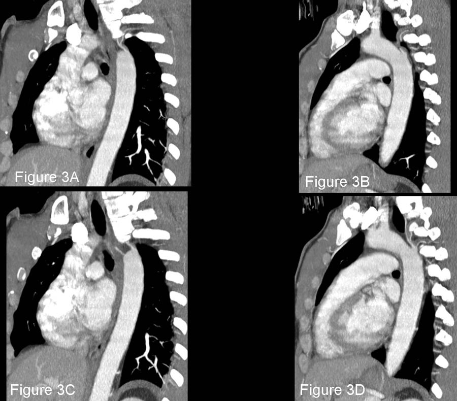
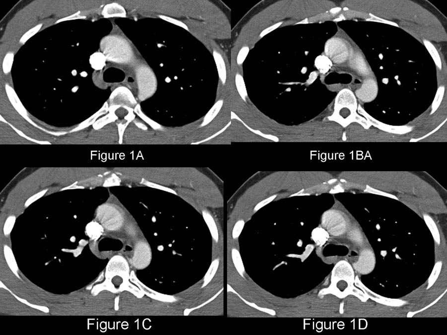
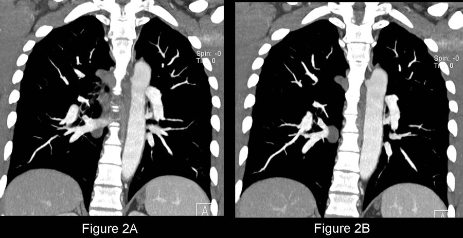
Contrast-enhanced chest CTA axial (Fig. 1A-1D), coronal (Fig. 2A-2B), and sagittal oblique (Fig. 3A-3D) images reveal a well-defined contour anomaly projecting posteromedially off the descending thoracic aorta. The contour anomaly is conical and triangular in nature and vessels emanate from its apex and course to the spaces (Fig. 1C; 2B; 3A-3D). The aorta is otherwise unremarkable.     Diagnosis: Prominent Right 3rd Intercostal-Bronchial Artery Trunk; No Vascular Injury

Discussion:

The infundibulum of the right 3rd intercostal artery is referred to by various terms including the right intercostal -bronchial trunk; superior intercostal; uppermost intercostal;  or highest thoracic intercostal artery. The infundibulum and its adjacent branches are often seen on Multiplanar chest CTA, MRA, and conventional transcatheter aortograms supplying the 2nd and 3rd intercostal spaces. The right infundibulum is usually more prominent than its counterpart and can mimic the appearance of an acute traumatic aortic injury or pseudoaneurysm. Other anatomic variants that can mimic acute vascular injuries include the brachiocephalic artery infundibulum, the aortic spindle, and a prominent ductus diverticulum (see Cardiopulmonary Pulmonary Imaging Correlation Conference Case of the Week from 9-24-2009).

Imaging Findings

CTA/ MRA / Aortography

  • Arise at the origin of the right 3rd intercostal artery (Fig. 1-3)
  • Conical, symmetrical, bulge (Fig. 1-3)
  • Smooth, uninterrupted margins (Fig. 1-3)
  • Vessel)s) emanate from its apex and course to the intercostal space(s) (Fig. 1C; 2B; 3A-3D)

Caveats

  • The characteristic appearance distinguishes this infundibulum from that of a true traumatic injury or pseudoaneurysm
  • Variations in the morphology of the infundibulum do occur; sometimes appearing more rounded than conical or triangular


Selected Readings

  • 1. Fisher RG, Sanchez-Torres M, Whigham CJ, Thomas JW. "Lumps" and "Bumps" that Mimic Acute Aortic and Brachiocephalic Vessel Injury. RadioGraphics 1997; 17: 825-834.
  • 2. Steenburg SD, Ravenel JG. Acute Traumatic Thoracic Aortic Injuries: Experience with 64-MDCT. AJR 2008; 191: 1564-1569.
  • 3. Scaglione M, Pinto A, Pinto F, et al. Role of Contrast-enhanced CT in the Evaluation of Acute Thoracic Aortic Injuries after Blunt Chest Trauma. Eur Radiol 2001; 11(12): 2444-2448.

Filed under: Radiology, Medicine/Pulmonary

Original case written by its authors at Virginia Commonwealth University and published at this address as part of a weekly teaching collection. Reproduced here as an archive.

Back to the case index