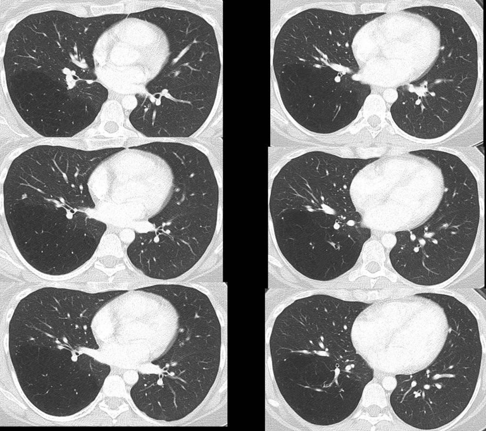
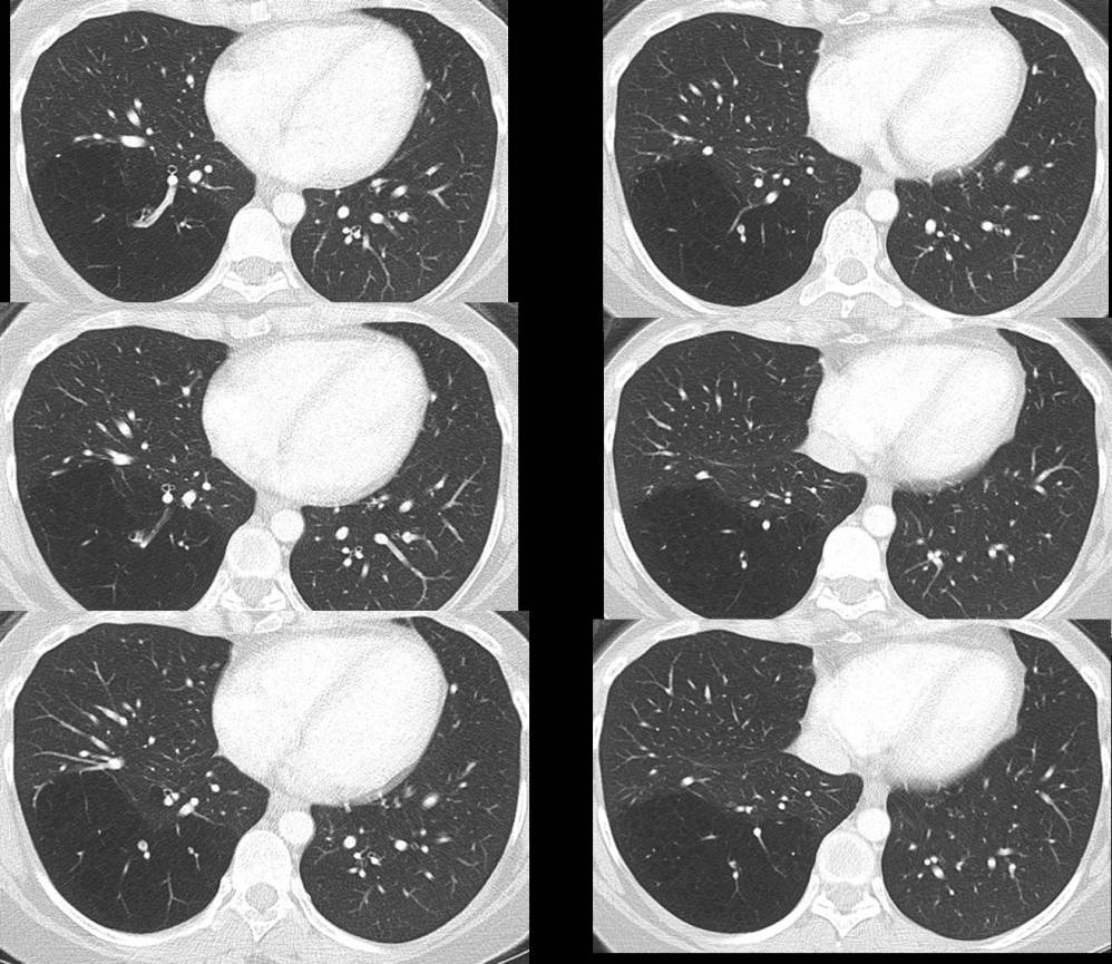
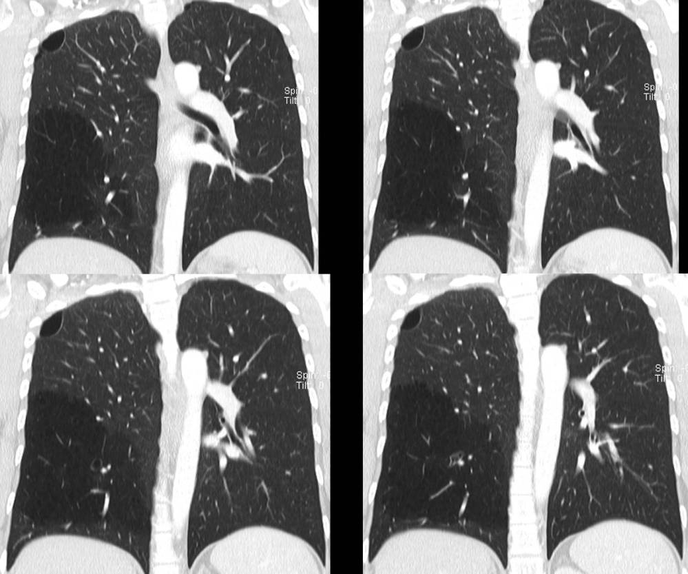
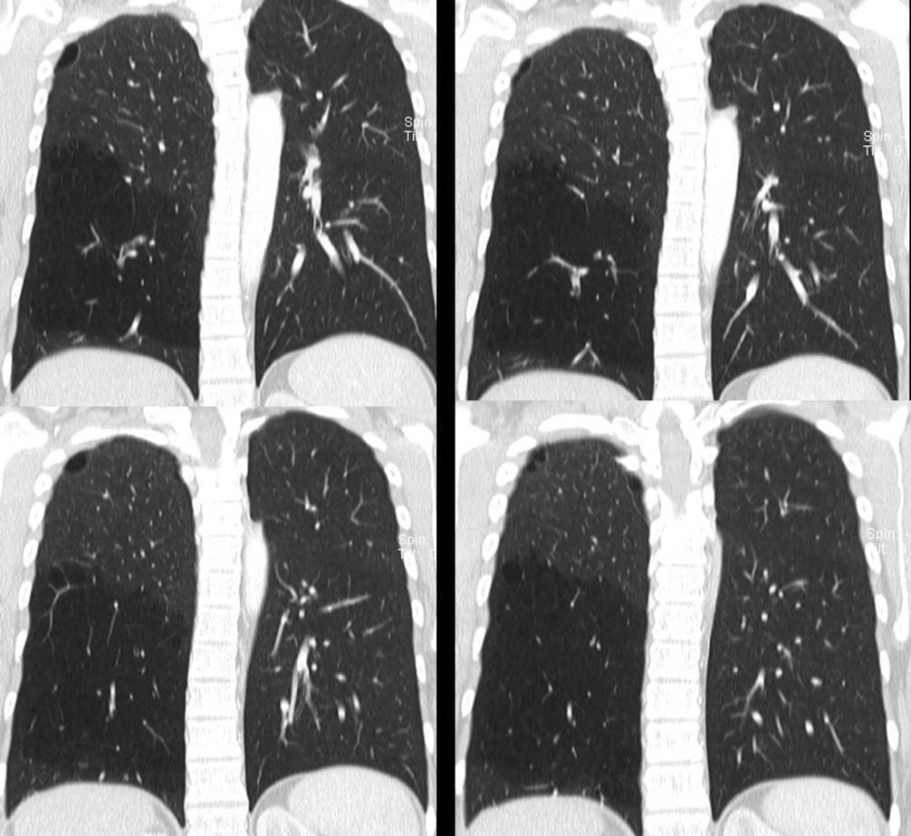
Axial (first group of 12 images) and coronal (second group of 8 images) CT (lung windows) demonstrate hyperinflation and relative oligemia of the right lower lobe, mild bronchial wall thickening and mild bronchiectasis with associated air trapping and subtle cystic formation. The right lower lobe lateral basal segmental bronchus abruptly stops between axial images 8 and 9 and coronal images 6 and 7. Scattered foci of mucoid impaction within the distally reconstituted segmental and subsegmental bronchi. Background of mild paraseptal emphysema. Diagnosis: Congenital bronchial atresia lateral basal segment of the right lower lobe
Differential Diagnosis
- Allergic Bronchopulmonary Aspergillosis (ABPA)
- Congenital Lobar Emphysema or Hyperinflation
- Focal Bronchiectasis with Mucous Plugging
- Intralobar Pulmonary Sequestration
- Mucous Plug Distal to Bronchial Obstruction
- Swyer-James Syndrome
Discussion
Bronchial atresia is a rare congenital anomaly characterized by focal narrowing or obliteration of lobar, segmental, or subsegmental bronchi resulting in an absence of communication between the segmental or subsegmental bronchus and the central airways. The left upper lobe bronchus is the most commonly affected (apicoposterior segment), followed by right upper and right middle lobes. Most cases are isolated and affect just one segment, but involvement of multiple segments and associated anomalies have also been reported.
Etiology
The exact pathogenesis of bronchial atresia is unknown; however, it is thought to result from an in-utero vascular insult that affects a small segment of the tracheobronchial tree with resultant interruption of the airway lumen and normal development of the distal airways. The obstructed lung may be fluid filled at birth and thus appear as a pulmonary mass. The surrounding alveoli may fail to develop normally but often remain aerated through collateral ventilation via pores of Kohn and canals of Lambert. The affected segment may then hyperinflate due to air-trapping. The post-atrietic bronchi are structurally normal, however may become ectatic and filled with mucous, resulting in a mucocele.
Clinical Findings
Patients are often asymptomatic and are diagnosed incidentally on chest radiography or CT acquired for unrelated reasons. Cough, dyspnea, bronchospasm, or symptoms of pulmonary infection may be found in up to 40% of patients. Men are affected twice as often as women.
Pathology
Gross
- Interrupted bronchial lumen and distended distal segmental bronchi
- Mucoid impaction
- Reduced size of affected pulmonary segment
Microscopic
- Reduced number of alveoli
- Alveolar over-distention
Radiologic Findings
Chest Radiography
- Focal pulmonary hyperinflation with increased volume and mediastinal shift towards the opposite side
- Elongated, tubular, or branching opacities representing mucocele distal to the obstructed segment
- Air trapping on expiration
- Air fluid level within mucocele
MDCT
- Triad of bronchocele, hyperlucent and hypoperfused lung
- Low attenuation branching, tubular, or rounded opacities with the distal lung (mucocele)
- Absence of contrast enhancement within bronchus or mucocele
- Hyperinflated lung surrounding mucocele
V/Q Scan
- Hypoperfusion of affected segment
- Delayed ventilation of affected segment with delayed washout (air trapping)
Treatment
- None for asymptomatic patients
- Surgical excision may be necessary in patients with chronic infection
Caveats
The imaging findings can be distinguished from Swyer-James syndrome by following the lateral basal segmental bronchus from its origin and observing its abrupt termination and the “over-inflation” of the affected lung.
Selected Readings
- Lange S, Walsh G. Radiology of Chest Diseases; Thieme, New York: 61.
- Lucaya J, Strife JL. Pulmonary Malformations Beyond the Neonatal Period In: Pediatric Chest Imaging: Chest Imaging in Infants and Children; Springer, New York: 93-110.
- Parker MS, Rosado-de-Christenson, Abbott GF. Tracheobronchial Anomalies In: Teaching Atlas of Chest Imaging 2006; Thieme, New York: 35-47.
- Web WR, Higgins CB. Congenital Bronchopulmonary Lesions In: Thoracic Imaging 2005; Lippincott Williams and Wilkins, Philadelphia: 1-29.
Original case written by its authors at Virginia Commonwealth University and published at this address as part of a weekly teaching collection. Reproduced here as an archive.