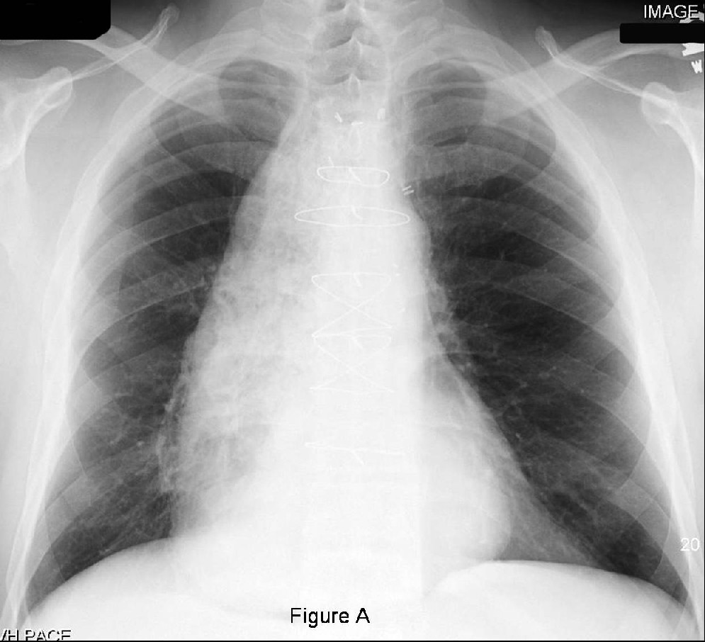
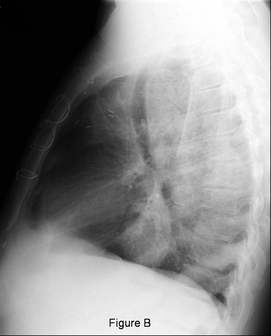
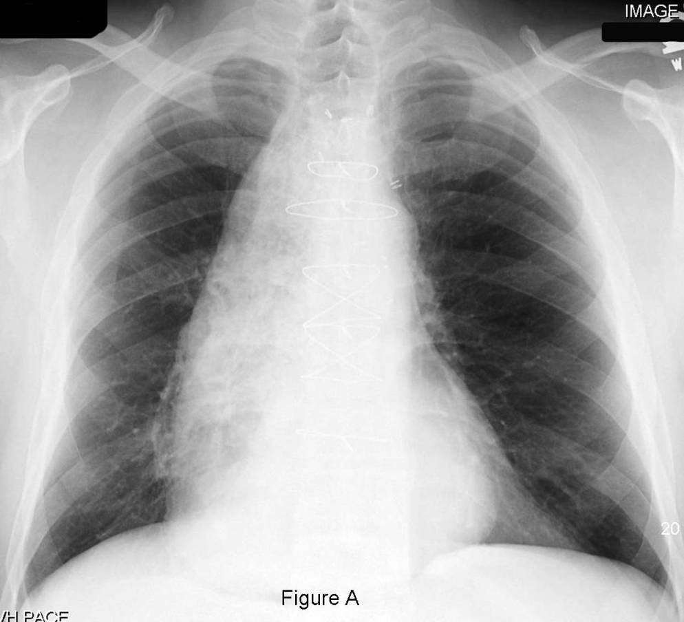
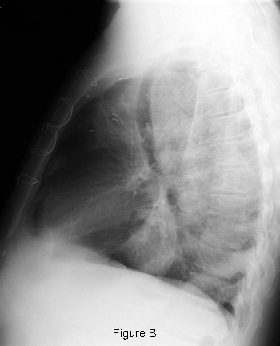
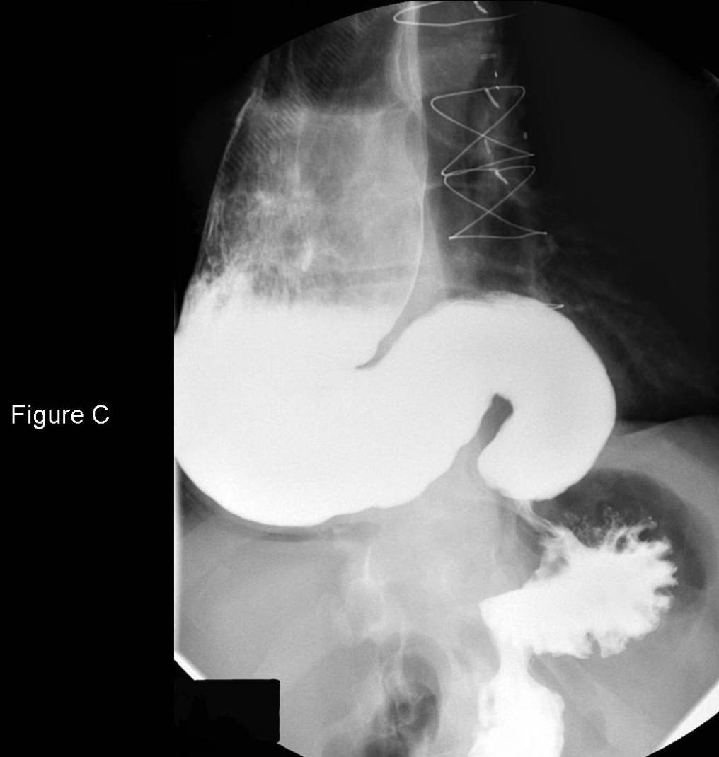
PA (Fig. A) and lateral (Fig. B) chest radiographs reveal evidence of an antecedent median sternotomy and coronary artery revascularization. Although the cardiac silhouette is not enlarged, there is an abnormal, eccentric, right-sided mediastinal mass present that does not silhouette the right heart border or ipsilateral hilum but does obliterate the azygos esophageal recess (Fig. A). The mass is of mixed density and extends from the level of the clavicles through and to the hiatus (Fig. A). The tracheal air column is displaced and bowed anteriorly by this middle mediastinal mass which obliterates the portions of the retrotracheal triangle, inferior hilar window, and retrocardiac clear space (Fig. B). Note the absence of visible gastric fundus air (Fig. A and Fig. B). Selected image from a double contrast esophogram demonstrates stasis and dilution of contrast and esophageal secretions in the dilated distal esophagus (Fig. C). Note the smooth, tapered narrowing or “bird-beak” appearance of the distal esophagus at the gastroesophageal junction (Fig. C). Diagnosis: Primary or idiopathic achalasia
Differential Diagnosis
- Secondary Achalasia from neoplasia
- Invasive gastric carcinoma
- Lymphoma
- Esophageal carcinoma
- Metastatic disease involving the gastroesophageal junction
- Chagas disease (Trypanosome cruzi)
- Strictures
- Extrinsic compression (e.g., hematoma)
Background
Discussion
Primary or idiopathic achalasia is a disease of the myenteric plexus (Auerbach plexus) of the esophagus in which peristalsis is markedly diminished or even absent. Secondary achalasia may simulate primary disease but is the result of underlying neoplasia.
Clinical Findings
Primary or idiopathic achalasia usually has its onset in early adulthood whereas secondary achalasia presents latter in life. Presenting symptoms may include dysphagia, regurgitation, halitosis from stasis or esophageal secretions, aspiration pneumonia, and esophageal candidiasis. Long-standing disease may also be complicated by an increased incidence of squamous cell carcinoma of the distal esophagus.
Imaging Findings
Chest Radiography
- Air-filled (“air esophagogram” sign) or fluid-filled tubular structure in the medial right hemithorax (Fig. A)
- Progressive esophageal dilatation and distention may obliterate normal mediastinal lines, stripes, and interfaces (e.g. azygos esophageal recess; inferior hilar window; retrocardiac clears space; retrotracheal triangle; etc) (Fig. A and Fig. B)
- Small or absent gastric fundus air (Fig. A and Fig. B)
- Anterior displacement and bowing of tracheal air column (Fig. B)
- Foci or air space disease, consolidation, basilar bronchiectasis (e.g., aspiration pneumonia; recurrent bouts of aspiration)
Esophography
- Disordered peristalsis; early in disease
- Diminished or absent peristalsis; latter in disease
- Dilution of barium contrast by retained esophageal secretions (Fig. C)
- Distended or dilated esophagus (Fig. C)
- +/- Megaesophagus
- Distal esophagus demonstrates a smooth, tapered narrowing caused by the contracted esophageal sphincter (“bird-beak” or “rat-tail” sign) (Fig. C)
- Gravity may partial overcome the decreased peristalsis and tightened sphincter allowing intermittent passage of small amounts of contrast media when patient is upright
- Little contrast passes through distal esophagus when patient is supine
Treatment
- Pneumatic dilatation
- Surgical myotomy
Selected Readings
- Mueller CF, Klecker RJ, King MA. Case 3. AJR 2000; 175: 867; 870-871.
- Parker MS, Rosado de-Christenson ML, Abbott GF. Achalasia. In: Teaching Atlas of Chest Imaging 2006; Thieme, New York: 691-694.
- Woodfield CA, Levine MS, Rubesin SE, Langlotz CP, Laufer I. Diagnosis of primary versus secondary achalasia: reassessment of clinical and radiographic criteria. AJR 2000; 175: 727-731.
Original case written by its authors at Virginia Commonwealth University and published at this address as part of a weekly teaching collection. Reproduced here as an archive.