Archived case 4 · Jun 16-Jun 23, 2009
Hepatopulmonary syndrome
37-yo man with liver disease admitted with GI bleeding and progressive hypoxia
Archived case 4 · Jun 16-Jun 23, 2009
37-yo man with liver disease admitted with GI bleeding and progressive hypoxia
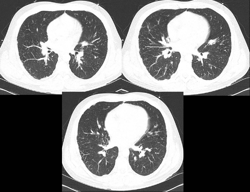
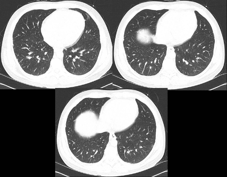
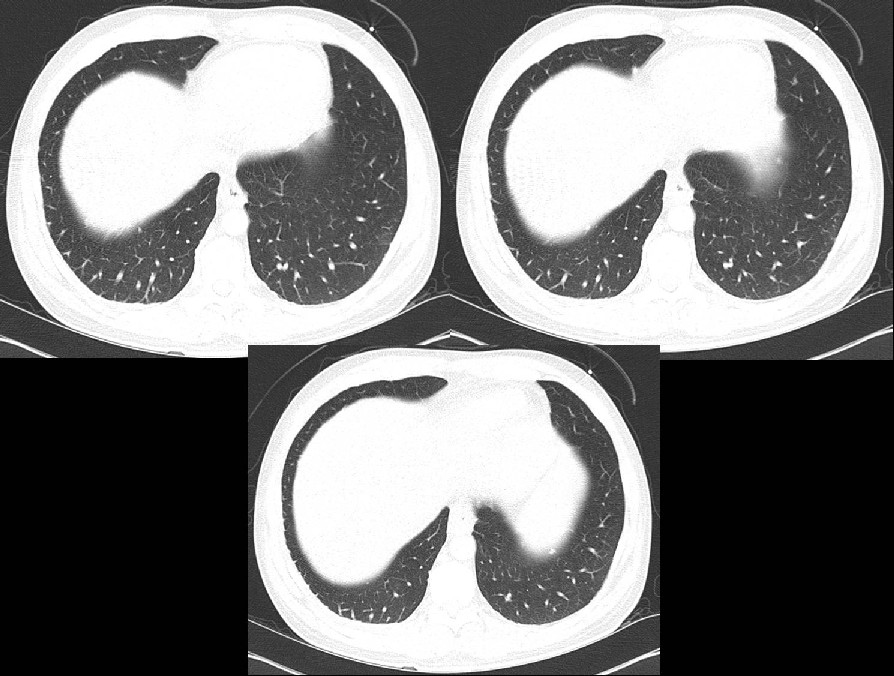
Hepatopulmonary syndrome
Differential Diagnosis
None
Discussion Background
Hepatopulmonary syndrome (HPS) is defined by the triad of liver disease, increased alveolar-arterial oxygen gradient ≥15mmHg while breathing room air, and intrapulmonary vascular dilatations. HPS develops in 15-20% of patients with cirrhosis. There are two types based on pulmonary angiography. Type I is the most common (86%) and manifests as distal vascular dilatation with juxtapleural telangiectasia. Type II (14%) is characterized by the formation of small, discrete peripheral arteriovenous malformations.
Etiology
It is postulated that HPS is due to the increased hepatic production or decreased hepatic clearance of vasodilators, particularly nitric oxide, by the impaired liver. This results in precapillary pulmonary artery dilatation, the formation of direct arteriovenous communications, and dilated pleural vessels. The vascular dilatation causes over perfusion of the lung relative to its ventilation, leading to a ventilation-perfusion mismatch and hypoxemia.
Clinical Findings
HPS manifests clinically as progressive dyspnea, cyanosis, spider nevi and digital clubbing in a patient with cirrhosis. Patients may also complain of platypnea and have orthodeoxia. Pulmonary artery pressures are usually normal or reduced.
Imaging Findings
Radiography
· Normal ; most common.
· Basilar, medium-sized nodular or reticular opacities.
Echosonography
· Bubble test is useful
o Intravenous microbubbles (>10 micrometers diameter) from agitated normal saline that are normally obstructed by pulmonary capillaries (normally <8-15 micrometers) rapidly transit the lung and appear in the left atrium within 7 heart beats.
Nuclear Scintigraphy
· Intravenous technetium-99m (99mTc)–labeled macroaggregated albumin may transit the lungs and appear in the brain, liver, and spleen.
· Distinction must be made from an underlying intracardiac right-to-left shunt.
Arteriography
· Type I
o Distal peripheral arterial dilatation.
o “Spidery” appearance of lower lobe peripheral juxtapleural vessels.
· Type II
o Small, discrete lower lobe pulmonary arteriovenous fistulae.
CT/HRCT
· Type I
o Dilatation of distal peripheral lower lobe pulmonary arteries that do not taper normally and extend out to the pleural surface.
o Increased number of visible peripheral pulmonary artery branches.
o Juxtapleural telangiectasia.
o Increased lower lobe segmental arterial diameter when compared with the adjacent bronchi.
· Type II
o Nodular dilatation of peripheral pulmonary vessels which are connected to a feeding artery and a draining vein.
· Intrathoracic portosystemic collateral vessels (e.g., coronary vein into esophageal or paraesophageal varices and cardiophrenic varices).
· Cirrhosis, hepatosplenomegaly, varices, ascites.
Treatment
· Currently no effective medical therapies but garlic powder and iloprost inhalation may be associated with some clinical improvement in the pre- and post-transplant period.
· Somatostatin (vasodilation inhibitor) is of modest benefit in some patients.
· Inhaled nitric oxide synthesis inhibitors may be an option in the future.
· Mainstay of therapy is supplemental oxygen
o Patients with PaO2 < 55mmHg, or PaO2 > 55mmHg with polycythemia, cor pulmonale, or cognitive impairment should receive 100% oxygen.
o If PaO2 increases to > 150mmHg and hypoxia is corrected, oxygen therapy should be continued (i.e., type I).
o If hypoxia is not corrected, or PaO2 < 150mmHg after receiving 100% oxygen, pulmonary angiography should be considered for potential embolotherapy (i.e., type II).
Prognosis
· Poor without treatment.
· Preoperative PaO2 £ 50mmHg alone or in combination with an isotopic shunt fraction ≥ 20% are the strongest predictors of postoperative mortality.
· Liver transplantation is the most effective therapy, correcting the syndrome in more than 80% of patients within 15 months of transplantation.
Caveats
· Do not confuse hepatopulmonary syndrome (shunting and V/Q mismatch due to vascular dilatation and arteriovenous malformations) with portopulmonary hypertension (development of pulmonary hypertension in a cirrhotic patient with portal hypertension).
· HPS is characterized by the triad of liver disease, increased A-a gradient, and intrapulmonary vascular dilatations. The liver dysfunction may not be severe.
Suggested Readings
Filed under: Radiology, Medicine/Pulmonary
Original case written by its authors at Virginia Commonwealth University and published at this address as part of a weekly teaching collection. Reproduced here as an archive.