Spontaneous pneumomediastinum complicating graft-versus-host disease (bronchiolitis obliterans)
Differential Diagnosis
Spontaneous Pneumomediastinum (sequelae of acutely elevated intrathoracic pressures)
- Illicit inhalational drug use (e.g., marijuana,; crack cocaine)
- Acute asthma exacerbation or other forms of chronic obstructive lung disease (e.g., bronchiolitis obliterans)
- Strong valsalva maneuvers (e.g., labor, parturition, weightlifting, straining).
- Severe upper respiratory tract infection (URI)
- Severe retching +/- esophageal rupture (Boerhaave syndrome)
- Severe cough
Traumatic Pneumomediastinum
- Barotrauma / mechanical ventilation
- Abrupt increase in intrathoracic and or intraabdominal pressure (e.g., crush injuries; motor vehicle collisions)
- Trauma or surgical disruption of the oropharyngeal, esophageal, or respiratory mucosa
- Subclavian vein catheterization
- Mediastinoscopy
Less common causes
- Interstitial lung disease
- Cystic lung disease
- Neoplasia of the aerodigestive tract
Background
Discussion
In this case, the patient developed bronchiolitis obliterans (constrictive bronchiolitis) as a manifestation of graft-versus-host disease (GVHD), leading to spontaneous pneumomediastinum. Bronchiolitis obliterans is part of the spectrum of chronic obstructive lung disorders that post transplant patients can develop, termed by some as “post transplant obstructive lung disease (PTOLD),” which has a high association with GVHD. The postulated mechanism for bronchiolitis obliterans in transplant recipients includes small airway damage secondary to medications, irradiation, ischemia, infections and autoimmune dysfunction. Other air-leak syndromes that can be manifest in GVHD and PTOLD include pneumothorax, pneumopericardium and subcutaneous air. Rupture of alveoli with subsequent air tracking along the peribronchovascular sheaths into the pulmonary hila and mediastinum is known as the Macklin effect.
Clinical Findings
Depending on the underlying etiology of the pneumomediastinum, affected patients may be asymptomatic or experience a wide range of clinical signs and symptoms including but not limited to: chest pain, cough, dyspnea, subcutaneous air if tracking occurs into the neck +/- Hamman sign (crunching of air over the precordium on physical exam)
Imaging Findings
Chest Radiography
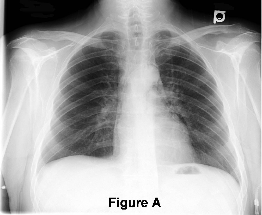
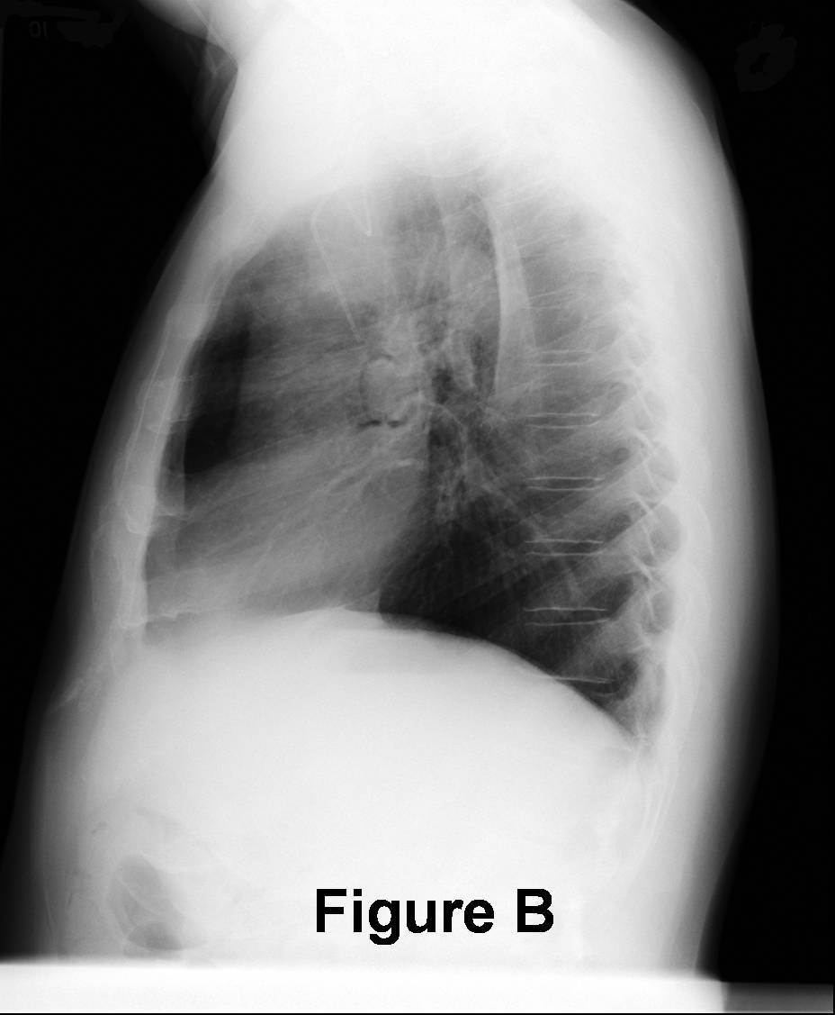
- Air outlining various mediastinal structures and or great vessels (Fig. A and B)
- “Double bronchial wall” sign (air on both sides of the airway)
- “Ring around the artery” sign (air dissecting around the pulmonary artery(ies) (Fig. A and B)
- “Naclerio’s V” sign (air outlining the descending thoracic aorta and extending between the parietal pleura and the medial left hemidiaphragm)
- “Continuous diaphragm” sign (lucency that appears to connect the right and left diaphragm beneath the cardiac silhouette)
- “Spinnaker sail” sign (pediatric cases; air elevates the thymus gland)
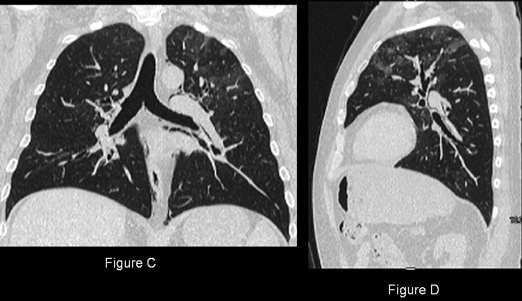
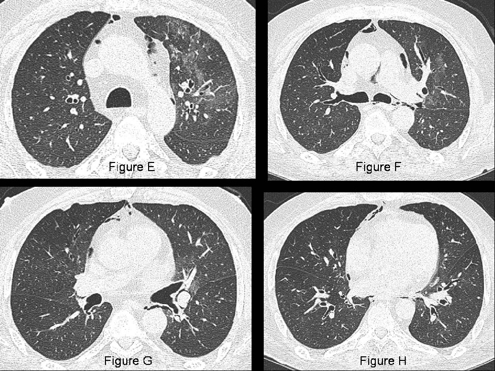
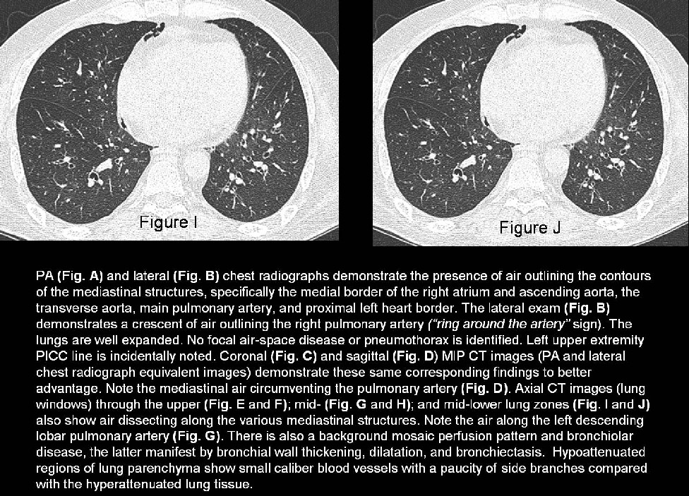
MDCT (Fig. C-J)
- Reveals the same imaging findings to better advantage
- May reveal underlying cause of the pneumomediastinum (e.g., tumor eroding through bronchus or esophagus)
- +/- concomitant pneumoperitoneum, pneumoretroperitoneum, pneumothorax
Treatment
- Pneumomediastinum itself rarely requires treatment except in cases of tension pneumomediastinum which may then require surgical decompression.
- Directed towards the underlying cause
Caveats
- Pneumoretroperitoneum and or pneumoperitoneum may dissect into the mediastinum and manifest as pneumomediastinum on chest radiography.
- Follow-up chest radiography should be obtained after 12-24 hours to asses for any progression of disease or unsuspected complications, such as pneumothorax.
- Black mach band may mimic pneumomediastinum and or pneumothorax. The
Mach band phenomenon is secondary to retinal inhibition with adjacent light and dark stimulation (e.g., soft tissue density adjacent to air density (left heart and lung) and will disappear if one edge is obscured (i.e. with a finger or hand).
Selected Readings
- Felson B. Chest Roentgenology. 1st ed. Philadelphia, PA: W.B. Saunders Company 1973.
- Galanis E, Litzow MR, Tefferi A, Scott JP. Spontaneous pneumomediastinum in a patient with bronchiolitis obliterans after bone marrow transplantation. Bone Marrow Transplant. 1997 Oct; 20(8):695-6.
- Gurney J, Stern E. Diagnostic Imaging for Radiology, STATdx. Accessed March 1, 2010.
- Iver V, Joshi A, Ryu J. Spontaneous Pneumomediastinum: Analysis of 62 Consecutive Adult Patients. Mayo Clinic Proceedings [serial online]. May 2009;84(5):417-421. Available from: Health Source: Nursing/Academic Edition, Ipswich, MA. Accessed March 1, 2010.
- Philit F, et al. Post-transplant obstructive lung disease ("bronchiolitis obliterans"): a clinical comparative study of bone marrow and lung transplant patients. Eur Respir J. 1995 Apr;8(4):551-8. Accessed March 3, 2010.
Original case written by its authors at Virginia Commonwealth University and published at this address as part of a weekly teaching collection. Reproduced here as an archive.