Methicillin-resistant staphylococcus aureus (MRSA) bacterial endocarditis with tricuspid valvular vegetations and septic emboli
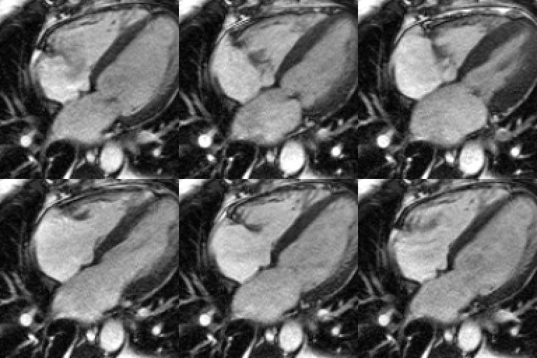
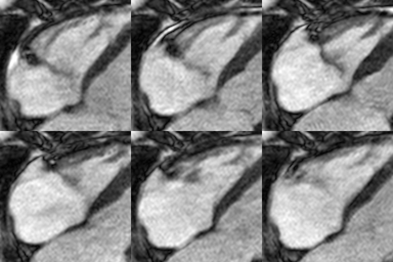
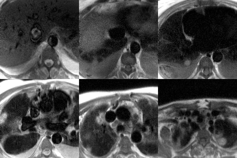
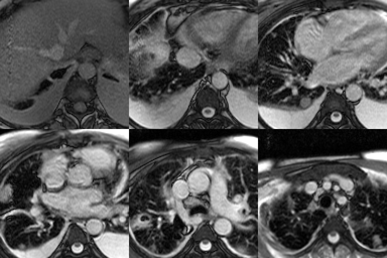
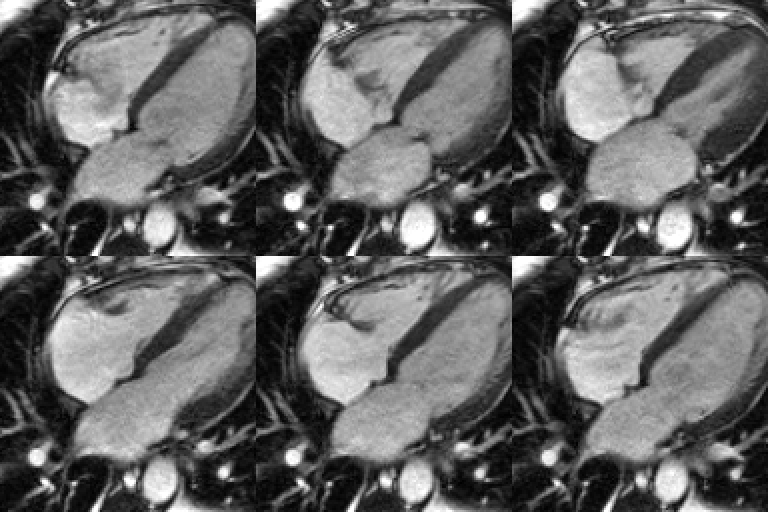
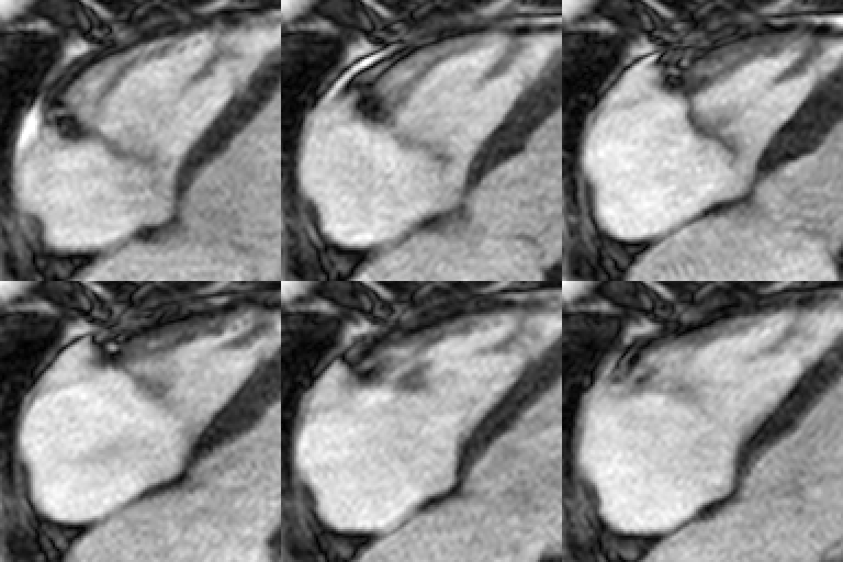
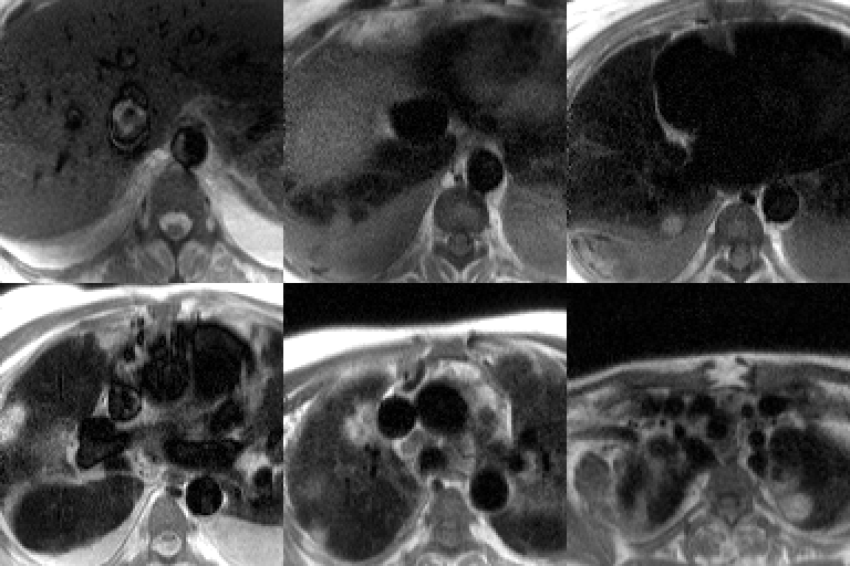
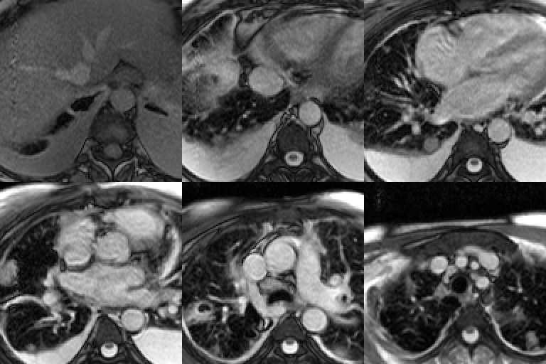
Four selected cardiac MRI sequences. Cine bright-blood 4-chamber (Fig. A) and small field-of-view (FOV) images targeted to the right heart and tricuspid valve (Fig. B) demonstrate irregular, lobulated, low-signal intensity thickening of both surfaces of the tricuspid valve. There is associated tricuspid regurgitation and right atrial dilatation. Axial HASTE (Fig. C) and short axis SSFP cine (Fig. D) images of the chest show multi-focal, variable-sized, predominantly peripheral and angiocentric nodular areas of parenchymal consolidation with varying degrees of cavitation. Note the bilateral pleural effusions and paratracheal lymphadenopathy. Diagnosis: Methicillin-resistant staphylococcus aureus (MRSA) bacterial endocarditis with tricuspid valvular vegetations and septic emboli
Differential Diagnosis
None
Discussion
Background
Endocarditis is an infection of the myocardial lining and or cardiac valves. Left untreated, endocarditis may be complicated by septic pulmonary embolism, cardiac dysrhythmias, valvular damage and or irreversible valvular destruction, and heart failure. Pulmonary septic emboli are frequently associated with intravenous drug use (IVDA) and endocarditis, especially with tricuspid valve infection and vegetation.
Etiology
Endocarditis is usually caused by hematogenous dissemination of various bacteria (e.g., Group A Streptococcus; staphylococcus sp) or fungi (Candida albicans). Cardiac valvular damage promotes the deposition of platelets and fibrin along the damaged valve’s surface. Bacteria or fungi may become trapped in these layers of platelets and fibrin forming “vegetations”. Foci of these valvular vegetations may subsequently dislodge and embolize systemically. Other sources of septic emboli include infected intravenous access sites and catheters and intra-abdominal abscesses. Risk factors for endocarditis include:
- Pre-existing cardiac valvular disease
- Artificial cardiac valve replacement
- Rheumatic heart disease with valvular damage
- Congenital heart disease (CHD)
- IVDA
- Hypertrophic obstructive or dilated cardiomyopathy (HOCM)
Clinical Findings
Persons with pre-existing heart and or valvular disease are more prone to develop endocarditis. Endocarditis also more commonly affects patients over 50-years of age, except in the setting of IVDA, in which case persons of any age may develop disease. Men are more often affected than women (2M: 1F). Patients with acute endocarditis may present with symptoms of fever, night sweats, myalgias, and fatigue. Chronic endocarditis may also be characterized by fatigue, night sweats, as well as arthralgias, weight loss, and heart failure. Physical signs of endocarditis include: Janeway lesions (i.e., erythematous spots on the soles and palms); Osler nodes (i.e., painful lesions on the distal phalanges); Roth spots (i.e., foci of retinal vascular hemorrhage); splinter hemorrhages under the finger nails; and new onset of a cardiac murmur or change in a pre-existing murmur.
Imaging Findings
Transesophageal echosonography (TEE)
- Gold standard for evaluating valvular vegetation
- Useful to assess for local complications, such as abscesses
- Visible vegetation suggests a worse prognosis
Conventional Radiography
- May be normal
- Cardiomegaly
- Cardiac decompensation
- Pulmonary complications such as septic emboli
- Typically multiple but can occur as a solitary lung nodule; mimic neoplasia
- Nodules generally evolve from a poorly, defined 1-2 cm diameter nodular opacity, to a moderately thick and irregular walled cavitary lesion
- When multiple septic pulmonary emboli are present; typically seen in the periphery of the lungs; usually in the mid - and lower lobes
- +/- Pleural effusion; parapneumonic effusion or empyema
CT
- Endocardial filling defect may be observed on contrast enhanced studies
- Valvular pathology may be detected on dedicated cardiac CTA
- Pulmonary complications such as septic emboli
- Solitary or multi-focal, variable-sized, predominantly peripheral and angiocentric nodular areas of parenchymal consolidation with varying degrees of cavitation
- Predilection for the mid - and lower lobes
MRI
- Excellent contrast resolution and high spatial resolution imaging of valvular structures very useful for establishing diagnosis (Fig. A-D)
- Vegetation intimately related to both surfaces of affected valve (Fig. A-D)
- Isolated or multi-valvular (e.g., TV and AV)
- Irregular and multi-lobulated (Fig. A-D)
- Low signal intensity before and after contrast administration; similar to thrombus (Fig. A-D)
- Valvular insufficiency or regurgitation (Fig. A and B)
- Pulmonary complications similar to those seen on CT (Fig. C and D)
Management
- Native valve endocarditis (NVE)
- Methicillin-susceptible staphylococci aureus. (MSSA)
- Oxacillin or cefazolin for 4 to 6 weeks.
- Methicillin-resistant staphylococci aureus (MRSA)
- Vancomycin; Gentamicin may be added
- Prosthetic valve endocarditis (PVE)
- May include MRSA or coagulase-negative staphylococci
- Vancomycin and Gentamicin may be used
- Rifampin also may be helpful in patients with prosthetic valves in addition to Vancomycin or Gentamicin
- Surgery
- Repair or replacement of damaged valve
Complications of endocarditis
- Myocardial infarction; pericarditis; cardiac dysrhythmias
- Cardiac valvular regurgitation
- Aortic root or myocardial abscesses
- Sinus of Valsalva aneurysm
- Arterial emboli; infarcts including mesenteric or splenic; mycotic aneurysms
- Heart failure
- Arthritis; myositis
- Glomerulonephritis; acute renal failure
- Cerebral vascular accidents (CVA)
Prognosis
- Dependent upon development of complications listed above
- Mortality rates in NVE; 16-27%
- Mortality rates in patients with PVE; higher
Caveats
- Endocarditis associated with IVDA
- Most commonly involves the tricuspid valve, followed by the aortic valve.
- S. aureus is the most common (<50% of cases) etiologic organism.
- Other causative organisms include streptococci sp, fungi, and gram-negative rods (e.g., Pseudomonas sp, Serratia species)
- MRSA accounts for an increasing portion of S. aureus infections; associated with previous hospitalizations and long-term addiction
- Coronary artery stents are not considered a predisposing risk factor for endocarditis
Selected Readings
- Chen JJ, Manning MA, Frazier AA, Jeudy J, White CS. CT angiography of the cardiac valves; normal, disease, and postoperative appearances. RadioGraphics 2009; 29(5):1393-1412.
- Grizzard JD, Judd RM, Kim RJ. Teaching File Cases. In: Cardiovascular MRI in Practice: A Teaching File Approach. Springer-Verlag, London Limited 2008; 124-125; 152-153.
- Rozenshtein A, Boxt LM. Computed tomography and magnetic resonance imaging of patients with valvular heart disease. J Thorac Imaging 2000; 15(4): 252-264.
- Vogel-Claussen J, Pannu H, Spevak PJ, Fishman EK, Bluemke DA. Cardiac valve assessment with MR imaging and 64-section multi-detector row CT. RadioGraphics 2006; 26(6):1769-1784.
Original case written by its authors at Virginia Commonwealth University and published at this address as part of a weekly teaching collection. Reproduced here as an archive.