Differential Diagnosis
- Acute presentation in symptomatic patient
- Incidental finding in asymptomatic patient
- Aortitis: normal interspersed segments between involved sites
- Periaortic lymphoma: thickened aortic wall with irregular external border
- Atheroma: irregular intraluminal surface
Background
Discussion
Aortic intramural hematoma (IMH) is often described as an atypical or “flapless” aortic dissection and is thought to represent either an early-stage limited dissection or thrombosis of the false lumen in dissection. The distinguishing feature is the absence of intimal disruption that characterizes classic aortic dissection. The Stanford system for classification of typical aortic dissection (AD) is applicable for the classification of IMH. That is, Stanford type A IMH involves the ascending aorta, with or without involvement of the descending aorta, and Stanford type B IMH involves the descending thoracic aorta, distal to the left subclavian artery origin. However, IMH exhibits a more variable natural history than classic AD, and may be characterized by periods of stabilization, regression, resolution, or progression to overt dissection. Type A IMH being more likely to progress to overt dissection than type B IMH. This is what was postulated to have occurred in this particular case.
Etiology
IMH results from rupture of the vasa vasorum and hemorrhage into the tunica media with resultant weakening of the aortic wall.
Clinical Findings
IMH is found in 5-20% of patients presenting with signs suggestive of acute (AD). 57% are classified as type A and 43% as type B. 94% of IMH are non-traumatic. Among IMH of a traumatic etiology, 75% occur in the setting of a motor vehicle collision. 61% of affected patients with IMH are men. The mean age at presentation is 63 years for men and 68 years for woman. Both IMH and AD have similar predisposing risk factors, the most common being hypertension, as well as clinical signs and symptoms (e.g., chest and or back pain). Less commonly, patients may experience syncope, hoarseness, anterior spinal syndrome or acute renal insufficiency. Additional clinical findings include ECG changes, aortic regurgitation, pericardial and pleural effusion.
Imaging Findings
MDCT (sensitivity / negative predictive value approach 100%)
Unenhanced CT
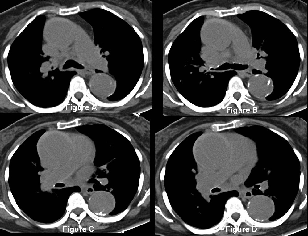
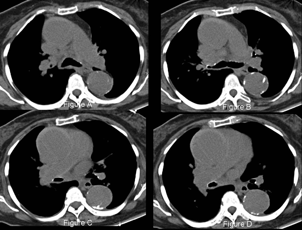
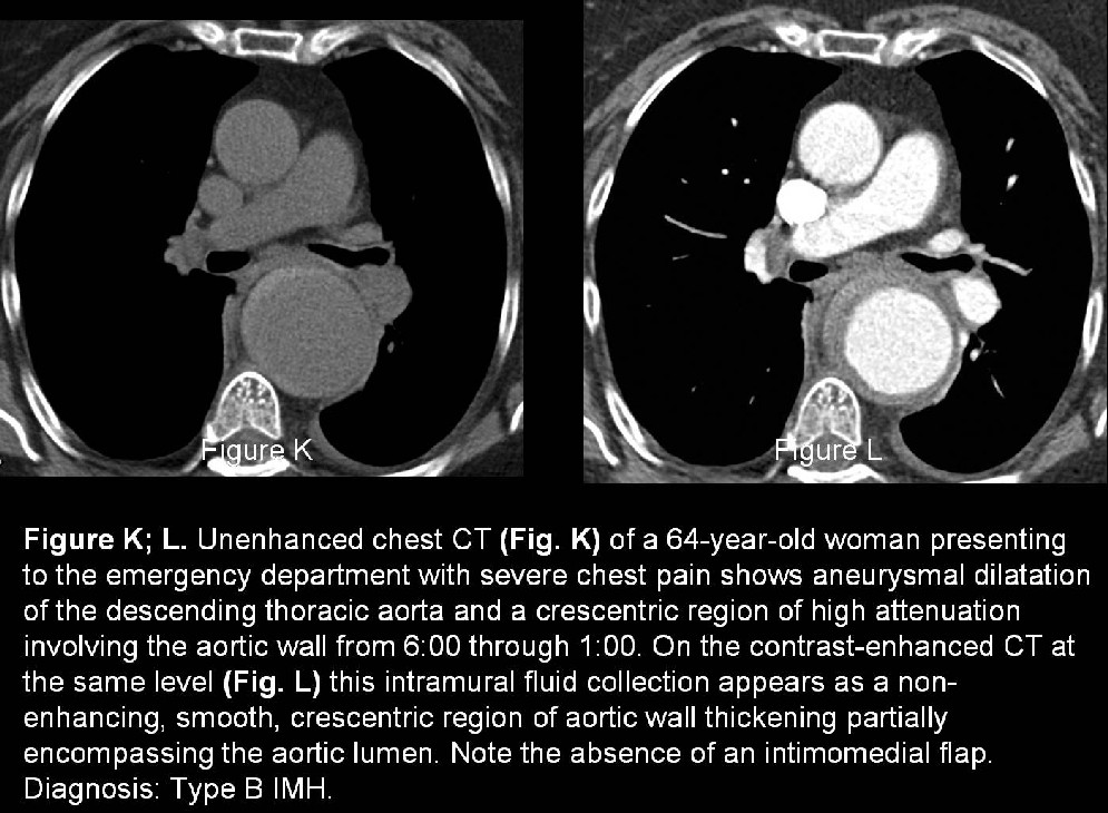
- Narrow window recommended (200 width; 40 level) for optimal depiction IMH (Fig. A-E; K)
- Enlarged aorta diameter (Fig. A-E; K)
- +/- Compression of aortic lumen
- Crescentric, eccentric, high attenuation region of aortic wall thickening (Fig. A-E; K)
- Inward displacement of intimal calcifications
- IMH: often appear semicircular or circular curvilinear
- AD: more often appear linear
Contrast-enhanced CT
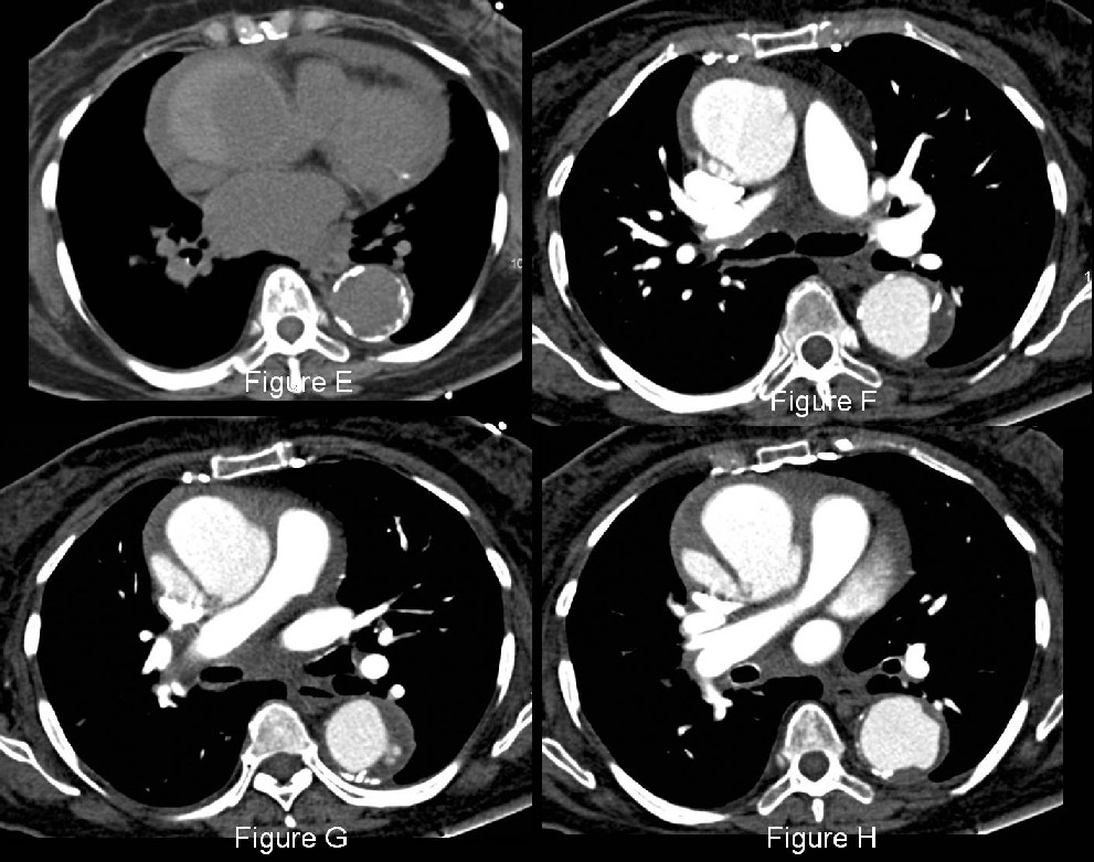
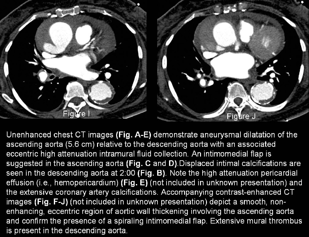
- Intramural fluid collection appears as a non-enhancing, smooth, crescentric region of aortic wall thickening (Fig. F-J; L)
- Intramural fluid collection extends partially or entirely around the opacified aortic lumen (Fig. F-J; L)
- No intimomedial flap, tear, or penetrating ulcer is present (Fig. L)
- Concomitant findings: mediastinal hematoma; pericardial or pleural effusion (Fig. J)
Transesophageal Echocardiography (TEE)
(Sensitivity 90-100%; specificity 91-100%)
- Focal aortic wall thickening
- Eccentric aortic lumen
- Displaced intimal calcifications
- Hypoechoic areas in aortic wall
- Limited evaluation of the aorta
- Severe atherosclerosis with focal wall thickening: False positive or equivocal results
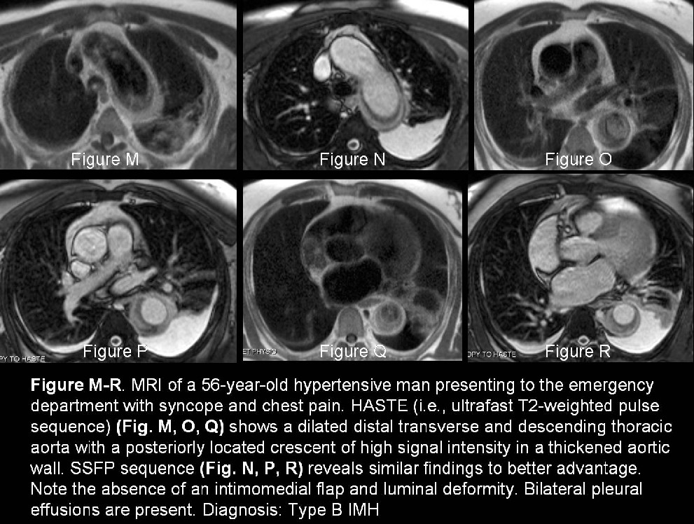
MRI (sensitivity 100%) (Fig. M-R)
- Aortic dilatation
- Crescentric intramural fluid collection (Fig. M-R)
- GRE (white blood sequences)
- Acute IMH (<7days): ↑ SI T2WI
- Subacute / Chronic IMH (≥7days): ↔ SI T2WI
- Spin-echo (black blood sequences)
- Acute IMH: ↔ SI T1WI secondary to oxyhemoglobin
- Subacute / Chronic IMH: ↑ SI T1WI secondary to methemoglobin
- Dynamic phase-contrast images: absence of flow in aortic wall
Angiography (sensitivity 83%)
- Limited usefulness for IMH
Complications
- New intimal tear (manifests as the formation of ulcer-like projections not present at time of initial diagnosis)
- Type A IMH >> Type B IMH
- More commonly occurs in ascending aorta and aortic arch
- 1/3 patients within first 3 months of follow-up imaging
- Saccular aneurysm formation at site of IMH
- Pseudoaneurysm
- Most commonly located in distal aortic arch
- Typically manifest 1week -7months after initial IMH diagnosis
- Enlarge at average rate of 1.3 cm / year; considerable potential for rupture
- Fusiform aneurysm formation at site of IMH
- True aneurysm
- More commonly located in descending aorta
- Progression to overt aortic dissection (Fig. F-J)
- Type A IMH >> Type B IMH
- Frequency type A IMH: 15-87.5%
- Imaging predictors of IMH progression to AD requiring surgical management
- Thicker IMH (16 mm vs 10.5 mm)
- Greater degree of luminal compression (ratio <0.75 of the minimum and maximum transverse diameters of aortic lumen at site of maximal IMH thickness)
- Maximal aortic diameter ≥ 50 mm strongest predictor (PPV 83%; NPV 100%)
- Predictive values of aortic regurgitation, mediastinal hematoma, pericardial and pleural effusion indeterminate
Management
- Type B IMH is conservative
- Type A IMH less well established
- Careful monitoring with regular follow-up imaging is mandatory even if the hematoma shows improvement or complete resolution because structural weakening of the aorta may result in delayed aneurysm formation or AD.
- No established guidelines on optimal frequency and longitudinal duration for surveillance. Some recommendations include
- Weekly CT for 1st month after diagnosis
- 2-3 CT exams during 1st year after diagnosis
Prognosis
- Morbidity and morality rate due to IMH similar to AD
- IMH mortality rate: 21%
- 1 week after symptoms: IMH attenuation similar to unopacified blood (NCCT)
- Follow-up imaging may reveal a decrease in IMH thickness within a few months and complete resolution within 1 year
Caveats
- The absence of an intimomedial flap, intimal tear, or penetrating ulcer is a prerequisite for the diagnosis of IMH.
- The radiologist should document the maximal aortic diameter, the maximal axial thickness of the IMH, and the minimum and maximum transverse diameters of the aortic lumen at the level of maximal IHM thickness. This data is useful for predicting the outcome of IMH.
Selected Readings
- Bluemke DA. Definitive diagnosis of intramural hematoma of the thoracic aorta with MR imaging. Radiology 1997; 204: 319–321.
- Chao CP, Walker TG, Kalva SP. Natural history and CT appearance of aortic intramural hematoma. RadioGraphics 2009; 29: 791-804.
- Fisher ER, Stern EJ, Godwin JD 2nd, Otto CM, Johnson JA. Acute aortic dissection: typical and atypical imaging features. RadioGraphics 1994; 14: 1263–1271.
- Gonsalves CF. The hyperattenuating crescent sign. Radiology 1999; 211: 37–38.
- Kaji S, Nishigami K, Akasaka T, et al. Prediction of progression or regression of type A aortic intramural hematoma by computed tomography. Circulation 1999; 100(19 suppl): II281–II286.
- Sawhney NS , DeMaria AN, Blanchard DG. Aortic intramural hematoma: an increasingly recognized and potentially fatal entity. Chest 2001; 120: 1340–1346.
- Yoshida S, Akiba H, Tamakawa M, et al. Thoracic involvement of type A aortic dissection and intramural hematoma: diagnostic accuracy—comparison of emergency helical CT and surgical findings. Radiology 2003; 228: 430–435.
Original case written by its authors at Virginia Commonwealth University and published at this address as part of a weekly teaching collection. Reproduced here as an archive.